


At this point, LASIK has become a quite common house hold term; however, what is actually involved in the process is far less well understood. LASIK is an acronym that stands for Laser Assisted in-situ Keratomileusis and refers to a refractive surgery for the correction of myopia, hyperopia, or astigmatism. As the name implies, lasers are integral to this process and contemporary refractive surgeries actually incorporate two different lasers into this process.
LASIK Procedure (three step process)
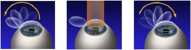
Step 1: Creation and Lifting of the “Flap”
The entire process of refractive surgery only involves manipulation and remolding of the cornea to compensate for an individual’s underlying glasses/contact lens prescription. The cornea is the clear tissue on the very front of the eye that overlays the colored part or iris and is a very thin tissue with an average thickness of about half a millimeter. Since this surgical procedure only involves the very front surface of the eye, no complex anesthesia is required. Eyes are simply numbed with topical anesthetic eye drops prior to surgery.
The laser used for the remodeling cannot simply be shot at the cornea and only works when used to laser the inside of the cornea (called the stroma). This restriction requires a very thin flap of cornea to be removed first. In the past, a small surgical bladed instrument called a microkeratome was employed for this task. However, the bladed technique had a tendency toward complications and often created flaps of varying thicknesses which resulted in unpredictable visual results.
These days a specific laser called the femtosecond laser is used for this task. The excellent video below shows the process of creating the LASIK flap using laser technology. With this method a soft suction ring is applied to the eye to hold the eye in place as well as flatten down the normally curved cornea. Once the cornea is flattened from the suction ring, the femtosecond laser forms a very thin layer of tightly packed bubbles at a specific depth in the cornea. These bubbles form a void inside the cornea allowing the flap to be pealed back and a hinge is left on one side. The suction needed for this process is slightly uncomfortable and causes a transient dimming of vision in the eye being treated, but as seen in the video, this process is very quick and the suction is quickly released. Additionally, once the flap is lifted, substantial blurring of vision will occur.
Forming the LASIK flap by using the femtosecond laser is widely considered a superior method for two reasons. First, the use of a laser allows a precise flap size and consistency which cuts down on a few commonly encountered flap complications like incomplete flaps, buttonholes (where parts of the cornea within the flap area are not cut), and free caps (where the corneal flap is completely cut off). Secondly, the laser consistently makes a flap of an extremely precise thickness of about 120-180 micros (depending on the surgeon’s preference) or roughly the thickness of three human hairs! The increased consistency of the flap gives surgeons more reproducible results in correcting the prescription and lessens the likelihood of retreatments or refractive eyewear needed after surgery.
Step 2: Laser Remolding of the Cornea
Once the flap has been made and lifted to expose the inside of the cornea (the stroma), a different laser called an eximer laser is then used to remold or remodel the cornea to compensate for an individual’s prescription. This laser ablates or essentially vaporizes the corneal tissue using extremely fast laser pulses (many hundred pulses per second) delivered in a finely controlled and step wise approach without damaging the tissue around it our any other part of the eye. This is a painless step and although very blurry after the flap is lifted, patients are able to fixate on a blurry light to allow the laser to work. Even with fixation, small movements of the eye during this step are unavoidable. To compensate for this, the laser has an incredibly advanced eye tracking system that follows the patient’s eye and makes corrections for small movements many thousands of times a second.
Even more amazing is that this entire complicated process of corneal remolding only takes 10-90 seconds depending on the amount and type prescription being corrected!
Below is a fascinating video of the eximer laser in action with actual video from the “patient’s perspective” captured by a smartphone.
Step 3: Repositioning the Flap
After the remolding (eximer) laser is finished, any debris from the procedure is washed and wiped away and the flap is carefully repositioned back in its original location without any wrinkles, bubbles, or irregularities. The flap is self-sealing and requires no additional fasteners like stitches.
Recovery
Following this outpatient procedure, patients are typically given antibiotic and anti-inflammatory eye drops to use for about a week and eye shields are required to be worn during sleep to prevent any accidental dislodging of the flap during the initial healing period. During the healing process, vision returns very quickly with most patients being cleared to drive and return to normal activities the day after surgery. Vision will continue to change slightly for several months as the eye heals, but typically vision is fairly stable after the first couple weeks. Retreatment or enhancement of the cornea is not expected but laser may need to be retreated if the prescription is not adequately corrected after the first procedure.
The better the surgeon, the lower the rate for retreatment, a great surgeon should have a retreatment rate of only 5-10%.
Complications
Severe complications of LASIK surgery are rare but can be sight threatening and include infections or immune reactions that can occur underneath the LASIK flap. These are rare but usually treatable.
There are some complications and side effects that are extremely common and include:
- Halos Around Lights – Far more common in patients who had surgery in the past. Halos occur from poor understanding of the treatment needed for the outer areas of the cornea and are more commonly noted in patients with larger pupils or in situations where the pupil dilates, like driving at night. It the past decade, surgeons have started paying much more attention to this and by measuring a patient’s unique curvature and corneal structure they are able to customize the laser treatment to maximize the optics of the new remolded cornea.
- Dry Eye – Another very common complication of LASIK. The cornea is highly enervated and sensitive and when the flap is created in the cornea these nerves are cut as well. These nerves let the brain know when the eye is drying. When this feedback loop is cut, the eyes don’t blink as much and don’t produce the necessary tears to properly wet the eye. Many of these nerves will regenerate to some degree by about 3 months after the procedure; however, some individuals have continued dryness for years after refractive surgery.
- Corneal Ectasia – Any screening done at a good surgical facility should include a thorough evaluation of the cornea to look for any hidden disease or potential complications. Even so, very infrequently LASIK can cause the cornea to loose its rigid structure and begin to bulge and warp. This is termed corneal ectasia and IS treatable with Specialty Contact Lenses. While this consequence is quite uncommon with LASIK, it is very common with older refractive surgeries like Radial Keratotomy or RK.
- Reading Glasses – It is very important to also note that LASIK surgery only corrects for distance vision. As our eyes age, the lenses of our eyes begin to harden and focus at near is lost. This process, known as Presbyopia or more commonly the “Curse of the 40’s”, happens REGARDLESS of laser surgery and reading glasses will still be needed for near tasks when this aging change occurs.
Other Refractive Procedures
LASIK is not the only option in the family of refractive surgeries. Other options include LASEK, PRK, ICL (an implantable lens), or clear lens exchange (cataract surgery without a cataract). These are all excellent options as well and may better suit the needs and desires of someone looking for freedom from glasses or contact lenses.
If you are curious about LASIK or any of these other options please feel free to ask for additional information at your next Complete Ocular Health Exam we would love to answer your questions!!