


The Anatomy of the Eye
Topics Covered:
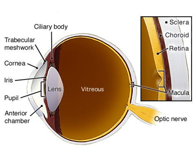
Vitreous | Sclera | Choroid | Retina | Macula | Optic Nerve | Central Retinal Artery | Central Retinal Vein
Vitreous
This is the jelly-like material that fills the inside of the eye. This jelly consists most of water, proteins, and collagen. Over time, these components break down and become sticky and form “clumps” in the jelly. As you move your eye, these clumps get shaken up and float around and many people see the shadows of these clumps floating through their vision. Commonly known as floaters, these are quite common and normally not a sign of any underlying problem unless significant new floaters are noted.
Sclera
Commonly known as the “white” of the eye, this tissue is a strong fibrous structure that covers most of the eyeball, gives the eye is structural strength, and protects the intra-ocular structures.
Choroid
The Choroid is a very thin membrane made up of blood vessels and sandwiched between the white of the eye (the sclera) and the retina. This membranous tissue consists mostly of blood vessels that nourish the outer part of the retina. The abnormal growth of vessels from this structure up into the retina accounts for the “wet” form of macular degeneration.
Retina
This thin multi-layered tissue lines the inside of the eye and actually captures the light that enters the eye and transmits that information to the brain. The retina has two types of light sensing cells: rods and cones. The rod cells are more abundant in the peripheral parts of the retina and are important for sensing light and dark changes as well as shapes and movement. The cones cells, by contrast, are heavily concentrated in the central retina, especially the macula, and sense colors as well as fine detail vision.
The proper health of the retina is absolutely critical to the function of the eye and is the site for many, many eye diseases and disorders.
Macula
This is the central part of the retina where cones are densely packed. It is the most sensitive part of the retina and is the only place out of which your eye can see perfect, 20/20 vision. Because of the heavy workload, the high concentration of cones cells, and the extensive vascular network surrounding this area, it is a common location for retinal problems to present including macular degeneration, diabetic eye disease, and many hereditary disorders.
Optic Nerve
This is the cable cord on the television so to speak. The optic nerve takes all the visual information collected by the retina photoreceptors and transmits that image back to the brain for processing and perception. Since the area only has nerve fibers and no photoreceptors (cones and rods), this is a natural blind spot that all humans have. The place where the optic nerve leaves the eye is called the optic disk and is the weakest part of the whole globe. Therefore, even though high eye pressure results from improper regulation of fluid at the front eye, it is the optic nerve at the back of the eye that gets damaged by increased eye pressure and glaucoma.
Central Retinal Artery
This artery enters through the optic nerve and supplies blood to the retina.
Central Retinal Vein
This is the main blood vessel that takes blood OUT of the inside of the eye and then meets up with other blood vessels to eventually take the blood back to the heart and lungs to pick up more oxygen. Veins look larger, redder, and flatter when viewed in the eye.
Click to read: “Anatomy of the Eye | Anterior Segment“