


Pellucid Marginal Degeneration or PMD is a bilateral (both eyes), non-inflammatory corneal disease characterized by severe inferior crescent shaped thinning.
Often times this condition is confused with Keratoconus and shares many of its characteristics and prognosis. However, most professionals agree that Pellucid Marginal Degeneration is a unique disease that presents specific challenges with specialty contact lenses.
What Causes Pellucid Marginal Degeneration?
Pellucid Marginal Degeneration appears to be completely idiopathic. In other words, there’s simply no reason why it develops. There is no gender, racial, ethnic, geographic, or socioeconomic links. There is also no links to other eye conditions. Nor does there appear to even be a genetic component.
This condition usually begins in the 2nd to 4th decade of life. Like Keratoconus, the thinning and bulging of the cornea is usually progressive and often times severe. Most of the time this condition is bilateral (both eyes), but it can occur in just one eye.
How Is Pellucid Marginal Degeneration Different from Keratoconus?
Both Pellucid Marginal Degeneration and Keratoconus are diseases of cornea ectasia, meaning corneal thinning and bulging. However, structurally, these two conditions cause corneal thinning and bulging in very different ways.
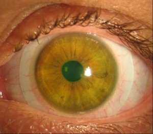
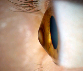 Keratoconus has a very characteristic cone shape bulge (above). While Pellucid Marginal Degeneration has a thin crescent shaped area of bulging at the bottom of the cornea (top of page).
This narrow band of peripheral thinning occurs from about 4 to 8 o’clock on cornea. Thinning in this area doesn’t result in a cone shaped area of bulging. Instead we see an overall sagging of the inferior cornea. This presentation is often referred to as a “beer belly” cornea.
Diagnosis With Corneal Topography
Under a microscope, these two conditions look quite similar.
Corneal Topography is the gold standard for diagnosing both Keratoconus and Pellucid Marginal Degeneration, as well as differentiating the two conditions.
Keratoconus has a very characteristic “cone” shaped area of thinning and bulging as seen with corneal topography.
By striking contrast, Corneal Topography with Pellucid Marginal Degeneration is very aptly described as “kissing doves” or the “crab claw.”
How is Pellucid Marginal Degeneration Treated?
Some severe complications with this condition are possible, such as acute corneal compromise (called Hydrops) or corneal rupture. Like keratoconus, if progression is noted, Cornea Cross Linking is appropriate to arrest the disease. Sometimes the thinning is so severe that a corneal transplant or Penetrating Keratoplasty (PKP) is indicated.
However, most of the time, this condition is simply treated with Specialty Contact Lenses.
 Since the area of thinning is so peripheral with Pellucid Marginal Degeneration, small diameter hard lenses like the Rose 2K are not effective. Their small size causes these lenses to slide too far down over the “beer belly.”
Instead, very large diameter hard lenses called Scleral Contact Lenses (seen above) are the standard of care. These unique lenses rest of the white of the eye and are very comfortable and well tolerated.
If you have any questions about Pellucid Marginal Degeneration or are in need of Specialty Contact Lenses including Scleral Contact Lenses, please contact our office.
Additional Links: