


Types of Keratoconus?
There are 4 main types of Keratoconus:
Forme Fruste, Nipple, Oval, and Globus.
Grading the types of keratoconus is largely based on where the area of bulging (or cone) is located on corneal topography.
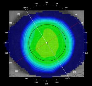
Essentially, corneal topography measures the elevation of the cornea similar to geographic topography that shows hills and valleys on a map. Topography is color coded with cooler colors (blues) being flatter and hotter colors (orange and reds) showing steepening from Keratoconus.
A normal topography (above) shows a very regular and uniform corneal surface without any significant steepening.
Forme Fruste Keratoconus
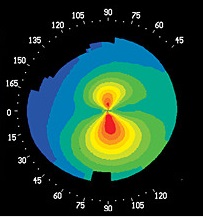 The mildest form of keratoconus is called Forme Fruste Keratoconus. Forme Fruste Keratoconus is sub-clinical and symptom free. Diagnosing Forme Fruste Keratoconus is really only possible through corneal topography.
With this form, the disease is present but is either very early/mild or has already stopped progressing completely. Above you can see the red inferior and asymmetrical steepening present on the topography.
This form of Keratoconus is extremely common and no treatment for this condition is indicated.
Typically glasses and soft contact lenses are all that is required for correction.
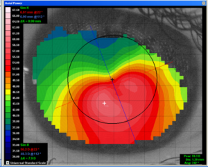
Nipple Cone
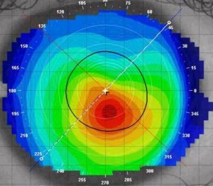
A Nipple Cone is a small isolated area of bulging at (or very near) the center of the cornea. Above you can see the relatively small but very pronounced area of bulging in red.
Although the area of corneal bulging is small, nipple cones often cause substantial distortion of vision because the area of corneal bulging is very near or in the pupil. The pupil location is denoted with the black circle.
This type of Keratoconus needs correction through Specialty Contacts. Depending on severity, the ideal treatment options could be specialty small diameter hard lenses, hybrid lenses, or scleral contact lenses.
Oval Cone
An Oval Cone is the most common form of keratoconus we encounter. Here the area of bulging is much larger and is mostly commonly seen inferior and temporally (toward the ear) on the cornea. Often there is a sagging appearance to the corneal on general observation.
While the area of thinning and bulging is down on the cornea, the corneal distortion does extend into the center part of the cornea and vision will be substantially decreased with glasses and soft contact lenses.
This type of Keratoconus would be be corrected through the use of Scleral Contact Lenses or possibly a Hybrid Contact Lens.
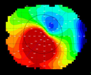
Globus Cone
A Globus Cone is simply defined as expansive corneal thinning and bulging affecting over 75% of the cornea (as seen above). This is the most severe form of Keratoconus but also the most uncommon. Globus Cones are more likely than other types of Keratoconus to require a corneal transplant called a Penetrating Keratoplasty (PKP).
Due to the extensive area of thinning, this type of keratoconus is likely only correctable through a large diameter Scleral Contact Lens.
Severity of Keratoconus
In addition the the type, Keratoconus can also be described has having a severity. The severity of the disease, along with the location of the steepening will correspond to the extend of the vision complaints and may also dictate what type of specialty contact lenses will work best.
Milder Keratoconus is often correctable with specially designed small diameter hard lenses like the RoseK 2K lens or hybrid contact lenses like the Ultrahealth Lens by SynergEyes. Moderate to severe Keratoconus are easier to correct with Scleral Contact Lens like the Zenlens by Alden Optical.
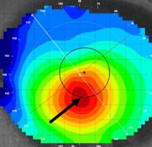
The severity of Keratoconus is graded by looking at the eye under the microscope and also running specialty testing to analyze the cornea to measure the steepest and thinnest areas (black arrow below).
Corneal Steepening
To grade Keratoconus by curvature, corneal topography is used to measure the absolute steepest part of the cornea at the height of the cone.
- Mild Keratoconus = steepest corneal curvature of ≤ 48.00D
- Moderate Keratoconus = steepest corneal curvature of 48.00D to 53.00D
- Advanced Keratoconus = steepest corneal curvature of ≥ 53.00D
Corneal Thinning
The average corneal thickness is about 555 μm (or a little over half a millimeter). Thinning at the bulging cone can lead to corneal break down, scarring, loss of vision, and additional complications that can lead to a Penetrating Keratoplasty (corneal transplant). Therefore, corneal thinning is often measured for monitoring progression and staging of the disease.
- Mild Keratoconus = Lowest corneal thickness of ≥ 500μm
- Moderate Keratoconus = Lowest corneal thickness of 300μm – 500μm
- Advanced Keratoconus = Lowest corneal thickness of ≤ 300μm