


In about a month, the Kentuckiana region is directly in the path of The Great American Solar Eclipse!!
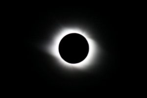
This solar eclipse will occur in our area on August 21st at 12:21 PM EST.
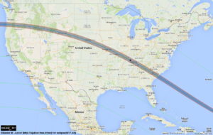
This eclipse will cross the entire continental US. While the eclipse will be visible from nearly every state, it will be especially spectacular in the Louisville area.
Here there is predicted to be a 90-95% partial eclipse, and if you head to southwestern Kentucky (around Hopkinsville) it will be a complete solar eclipse for about 2 minutes!
These events area extremely rare with the last total eclipse occurring here back in February 26th, 1979!!
While this once in a life time event is amazing, it is also EXTREMELY DANGEROUS!
It is absolutely critical that this miracle of nature be viewed through an approved device.
These devices include:
- ISO Certified eclipse glasses (certification 12312-2:2015)
- Welders Shield or Filter with shade #12-14
- Viewed indirectly with a Pinhole Camera – A great project for kids! (NASA project link)
Wearing sunglasses or even ‘taking a quick peek’ can cause irreparable damage.
Don’t Risk It!!
Solar Retinopathy
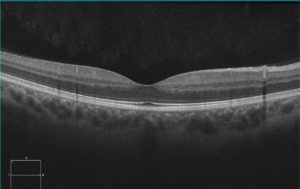
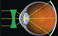
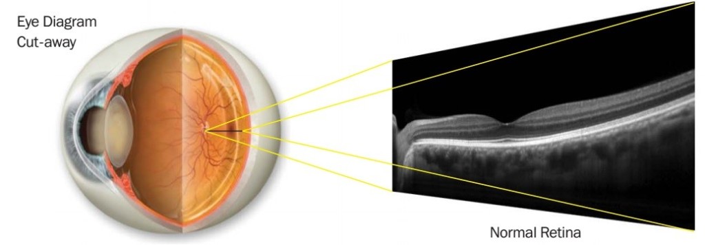
The eye focus light that enters the eye back onto the retina. In particular, the light is focused most strongly onto the macula, which is the center part of the vision (normal imaging of macula above).
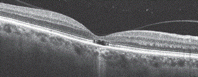
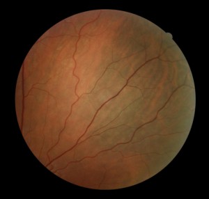
However, when high intensity, direct sunlight is focused on the macula, it heats up the pigmented layer underneath the retina. This layer gets so hot from the high intensity light that it actually burns the cells that sense light in the center part of the vision (see above).
When this area is damaged, vision will be decreased and you may noticed a blind spot in the center of your vision. Sometimes this condition improves over time but often the vision does not completely return and permanent vision loss occurs.
If you would like to know if the eclipse glasses you have are safe, please email or stop by our office and we’d be happy to talk with you further.
Be Safe and Enjoy the Eclipse!!
Dr. Wolf
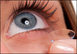 Today on Eye to the Future we will be discussing a type of contact lens you probably haven’t heard of before, but one that has helped many of our patients live fuller, happier, and more visually productive lives – Scleral Contact Lens.
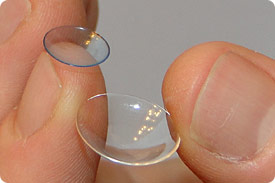 A newer type of hard lens for eye disease is the scleral contact lens.
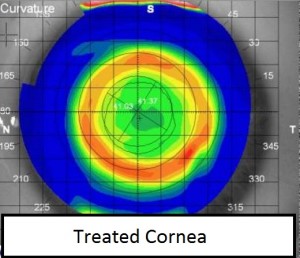
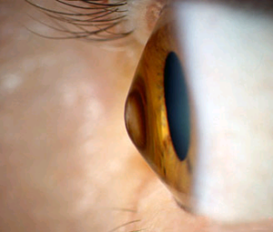 The cornea is the clear front surface of the eye. Normally, the cornea has a round regular shape, like a ball. Other times the cornea is more egg or football shaped. This is called an astigmatism and is also very normal.
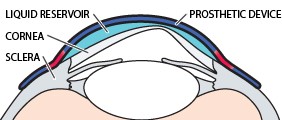 The cornea is the first surface that light goes through. The bulging irregular surface from ectasia heavily distorts the image going through it.
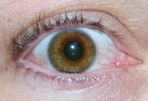
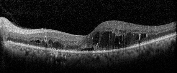
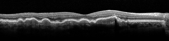
 Patients with severe dry eye require copious amounts of lubrication to achieve comfort and provide sustained, acceptable vision.
 Deli-Style Kale Salad
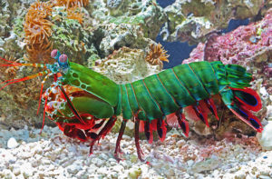 Think you see pretty well?
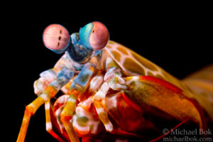 The mantis shrimp has two eyes, same as us, but that is where the similarities end.
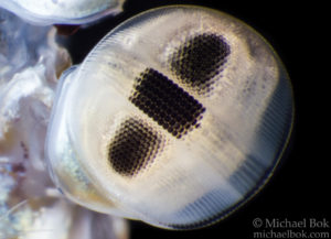 The mantis shrimp’s eye is separated into three different sections. The upper and lower hemispheres are used mostly for sensing motion and forms. The central area is called the midband and contains 6 rows of specialized clumps of photoreceptors.
 Superlatives don’t stop there with these little critters!
 Since the eye grows front-to-back, the whole eye isn’t growing, just the parts around the outside of the eye in the peripheral retina. If light entering the eye doesn’t fall properly on this part of the retina (shown left), it stimulates the eye to grow backwards to focus the light. Unfortunately, this growth causes the light falling in the center part of vision to be out of focus resulting in fuzzy distance vision.
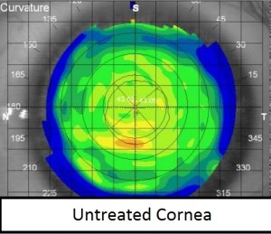
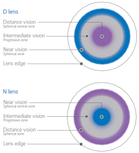 The newest, and most exciting advancement in myopia progression control is through the use of Bifocal Contact Lenses.
  Especially rich in: eye-healthy omega-3s DHA and EPA.
 If I had a dollar everytime I heard that from patients… well I sure wouldn’t be rich, but it actually does happen a lot!
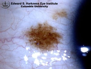
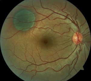
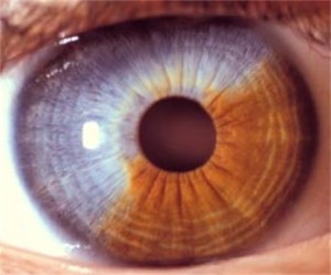 The human eye forms initially as an in-pouch of embryonic skin cells and since it also has melanocytes, you can also get these freckles or nevi on any part of the eye. Here’s one on the white of the eye called a conjunctival nevus, as well as a large iris nevus. These moles are easily seen in the mirror, what comes as a shock are the nevi inside the eye or choroidal nevus.
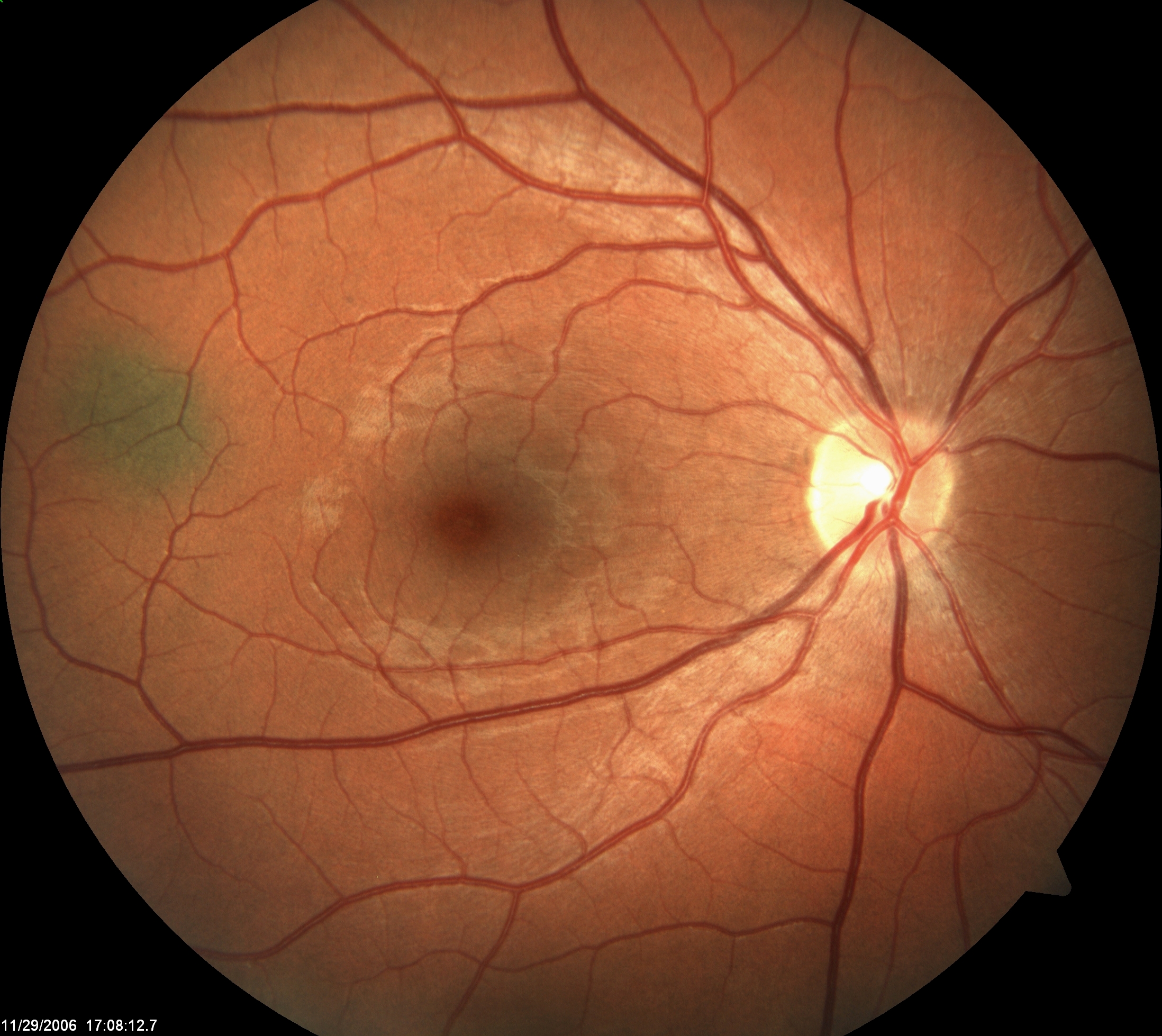
 Choroidal nevus inside the eye can come in all shapes and sizes as well. While all nevi are by definition “normal,” they can be dark, light, small, or very, very large. The first is a relatively small mole about 1.5 mm in size, the second photo is part of a much larger that covers about a quarter of the inside of this persons eye!