Back to School: Don’t forget the Eye Check!

PediatricEyeCareWith school back in full swing, Precision Family Eye Care and the Eye to the Future blog want to take a moment this week to impress upon all parents the importance of getting routine vision exams for all school age children.

School and Pediatrician screenings are simply not adequate, and unlike adults, children can’t articulate their vision problems because they don’t understand that the way they see the world isn’t “normal.”  Uncorrected prescription (like nearsighted, farsighted, and astigmatism) as well as eye alignment and focusing problems, and even ocular diseases can all inhibit the ability of your child to use their eyes properly.  This leads to disrupted vision and potentially vision threatening conditions being simply overlooked or even causing learning problems and leading to a diagnosis of LD (Learning Disorders), ADHD or even dyslexia.

The learning environment today is flush with visual tasks: smart boards, iPads, digital learning, computer work, homework, test taking, etc.  It shouldn’t surprise us then, that learning experts estimate that roughly 80% of all learning is dependent upon the sense of sight and unfortunately, at least 25% of school aged children have a visual impairment that may prevent a child from reaching their full academic potential.

Please make sure that your child has the proper tools for academic success this year by scheduling a comprehensive exam with your eye doctor today!

Patient Question: “How do animals see in the dark?”

As is often the case, most of the ideas for this blog come from actual questions asked by patients.  On Eye to the Future today we will be answering one of these questions I received just last week, “How do animals see in the dark?”

GREAT QUESTION!

How exactly does my dog and cat run scot-free through a dark room but I stub my toes and step painfully on toys?

The answer: it’s all in the design of the eye.

Thanks for stopping by, next week on Eye to the Future we will be discussing… kidding of course.

To answer this question fully, there are actually three major anatomical differences between the human eye and animal eyes capable of great night vision.

Rods versus Cones

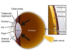
CataractsDiagramAs most are aware, the retina is the covering of the inside of the eyeball.  Although this thin tissue is only about 3 human hairs thick, it is essentially the ‘film in the camera’ and is responsible for sensing the light that enters the eye and transmits that signal back to the brain where it is processed.  The actual cells that sense the light are called photoreceptors and there are two main types: rods and cones.  Although they both are important in seeing, they do so very, very differently.

Cones are responsible for color vision and high acuity vision however these cells only function in bright light.  Rod cells, by contrast, are much less sensitive to colors and fine details but are able to detect light and function in much lower light situations.

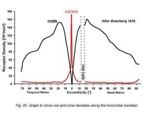
In humans, 90% of our cones cells are located in the central part of our vision which allows us vivid, clear, and colorful central vision, but general poor vision under scotopic (dark) conditions because of a lack of rods in our central vision (graph at right).  Rod and Cone DistributionBy contrast, nocturnal animals have retinas composed mostly or even entirely of rods cells, especially in their central vision.  This affords nocturnal animals great night vision; however, their lack of cone photoreceptors leaves these animals with little to no color vision and an overall decrease in clarity compared to humans.

Bigger is Better

Since nocturnal animals cannot benefit from the crisp vision that cone cells allow, they require a much larger image to focus on their retinas.  The easiest way to accomplish this is with a larger eye and larger eye structures (like a stronger focusing cornea and lens as well as a larger pupil to let in more light).  The human eye is about 24 mm (about 1 inch) in diameter.  The eyes of nocturnal animals are substantially larger even through the animal itself can be much, much smaller.  The owl is a perfect example of this since it has an eye that is about the size of a human eye but gives enough visual information to allow the owl to be a successful night bird of prey.  However if we are talking size, the most dramatic example and clear winner of the “Biggest Eye on the Planet” award goes to the giant squid which is able to visualize prey in the pitch black of the ocean’s depths with an eye that’s the size of a dinner plate!!

Tapedum Lucidum

Although larger eyes and specialized retinal cells are certainly useful, nocturnal animals have one final trick up their sleeves to help with night vision.  The retina in all animals is a clear, see through tissue.  The reason for this is because the rod and cone cells that actually sense the light are at the back of the retina, so the layers above need to be clear to let light get through.  Now in a human eye, once the light goes through the retina it hits the retinal pigmented epithelium or RPE.  This thin layer of cells is highly pigmented and absorbs excess light that gets through the retina to reduce glare/scattered light and improve overall clarity of vision.

However, many nocturnal animals like deer, raccoons, cows, horses, and even dogs and cats, have developed a specialized RPE behind the retina called the Tapedum Lucidum that does the exact opposite.  Instead of being highly pigmented to absorb the light that the retina does not collect, the Tapedum Lucidum is a highly reflective tissue, like a mirror, that actually bounces the light back through the retina for a second chance to be detected.  This adaptation effectively doubles the chance that even very low amounts of light will be detected by the retina.  Reflections off of this tin foil like Tapedum Lucidum is also what causes the brilliant white reflections from the eyes of nocturnal creatures, like a deer in headlights or even a photo of the family pet.

Tapedum LucidumWe hope you have enjoyed this long answer to a short question.  Next time on Eye to the Future, we will be discussing polarized lens.  How they work, how they’re made, and why they are so, so useful for clarity and vision.
I’ll leave you with a picture of our sweet boy, Remy, taking a rest from reeking havoc and showing off his Tapedum Lucidums.

See you next time on Eye to the Future.

STUDY: Age Related Eye Disease Study (AREDS 1 & 2)

On occasion, there are landmark studies regarding the eyes that everyone should know about.  Today on Eye to the Future we will be discussing two such studies, by far the largest and most important studies on preventing the progression of the aging condition Macular Degeneration.  (For those not interested in the science, the results and take home points are summarized at the bottom of this post.)

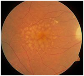
Dry Macular DegenerationAge Related Macular Degeneration (ARMD or simply AMD) is the leading cause of new blindness in the United States for individuals over 65 years old.  This condition is not a new problem, however, for many decades the mechanism for why this damage occurred was unknown and treatment options for this condition were woefully limited and relatively ineffective.  Thankfully, in the last decade and a half, some genius innovations in treatment, as well as some landmark studies on prevention, have given individuals afflicted with this blinding disease a fighting chance at retaining vision throughout their lives.

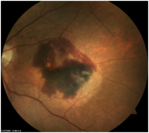
Wet Macular DegenerationMacular Degeneration can be classified into two categories Dry and Wet (for an in-depth discussion on the difference between the two, please view our website for a thorough discussion).  Dry Macular Degeneration (above) always comes first and by itself can cause vision loss, however, more often it is the 10-15% of patients that convert to the Wet form (with its associated bleeding and scarring) that suffer severe permanent damage.  Therefore, researchers set out to look for ways at preventing or at least slowing down the conversion to this more aggressively blinding Wet form (right).  The research culminated in the Age Related Eye Disease Study (AREDS) that was first published in October of 2001, and overnight, changed the way eye care practitioners approached macular degeneration.

AREDS 

It had been hinted at in smaller previous studies that antioxidants as well as the trace element Zinc appeared to be helpful for macular degeneration patients.  As a direct result, many patients starting taking high doses of these vitamins and minerals without any set dosing amounts, schedules, or any singular study that ensured this practice was even effective… or safe for that matter!!  It became clear that a definitive study was needed and the National Eye Institute, one of the government’s Institutes of Health, brought onboard a decorated panel of eye doctors, biochemists, and nutritionists who came together and concocted the follow mixture of supplements.

Vitamins

o   Beta-Carotene (15mg) which is converted in the body to Vitamin A

o   Vitamin C (500 mg)

o   Vitamin E (400 IU)

Trace Minerals

o   Zinc (80 mg), which was shown to be effective in earlier studies

o   Copper (2 mg) to prevent anemia while taking high doses of Zinc.

Over 3500 patients with various amounts of Macular Degeneration took this supplement for a minimum of 5 years.  Once all the data was tabulated, the most encouraging results were for the patients who needed it most with intermediate to advanced Macular Degeneration.  The results for patients in this category are outlined below.

Vitamins Alone

  • Reduced the risk of advanced ARMD by 17%
  • Reduced vision loss by 10%
Trace Minerals Alone

  • Reduced the risk of advanced ARMD by 21%
  • Reduced vision loss by 11%
Vitamins AND Trace Minerals

  • Reduced the risk of advanced ARMD by 25%
  • Reduced vision loss by 19%

Once it was shown that this nutritional supplement formula of both vitamins and trace minerals decreased advanced Macular Degeneration by 25% and vision loss by nearly 20%, many different nutraceutical companies began reproducing the AREDS Formula with brand names that you have likely heard of or have seen in stores like I-Caps™, PreserVision™, Ocuvite™, et al.

AREDS 2

Not long after the AREDS was published, the researchers behind the AREDS study went back to work on an even larger more detailed study using the information gleaned from the first study.  The results of the AREDS 2 were just released in 2013.  This improved study adjusted two of the past components of the AREDS formula and also added a third.

  1. Beta-Carotene:  Very high doses of Beta-Carotene/Vitamin A supplements have been linked to an increased risk of Lung Cancer in current or former smokers.  Since smoking in-and-of-itself is already a significant risk factor for developing Macular Degeneration, beta-carotene was removed completely for safety reasons and replaced with two other carotenoids: Lutein (10 mg) and Zeaxanthin (2 mg) which were deemed a safer and equally effective substitute for Vitamin A.
  2. Zinc:  It was suspected that high dosages of Zinc are linked to digestive problems.  Reducing Zinc (to 25mg)  showed the same benefit as the AREDS formula, but removing Zinc from the formula completely was detrimental.
  3. Omega 3 fatty acids (fish oils):  These compounds are a known anti-inflammatory and antioxidant.  Researchers added this compound to the AREDS formula in hopes of additional reduction in risk, however, Omega 3’s were shown to have no effect on Macular Degeneration progression.

AREDS 1 and AREDS 2: Take Home Points

  • Any patient with confirmed macular degeneration will benefit from eye specific supplements, but these supplements are most helpful for patients with moderate to advanced Macular Degeneration.
  • These supplements should be taken IN ADDITION to any multivitamins, not as a substitute (9 out 10 patients in the AREDS 2 study were also taking a concurrent multivitamin).  If only one supplement is desired, there are formulations of these supplements that combine the AREDS formula with a multivitamin for simplification.
  • The original AREDS formula is fine for many, if not most patients.
  • Any smoker or former smoker should avoid beta-carotene supplements completely and use only the AREDS 2 formula with Lutein and Zeaxanthin instead.
  • Omega 3 fatty acids (fish oils) although wonderful for the body and the eyes, did not have a meaningful impact on Macular Degeneration.
  • These studies ONLY found benefit for patients with diagnosed Macular Degeneration.  These supplements are not designed, and should not be taken for “routine” eye health or even for patients with a family history of Macular Degeneration.

We hope you found this discussion interesting.  As always, if you have any questions or have ideas for future topics feel free to contact us and ask.  We love questions!

Eye to the Future next week will be a fun one… but I’ll let it be a surprise.


“Pink Eye” Myths: What it is and What it’s not.

Well, it’s back to school time and that means it’s also time for some germs to be spread and flus to be caught.  This time on Eye to the Future we will be talking about the eye’s contribution to the germ problem with Pink Eye.  The term Pink Eye gets batted around quite a bit in most people’s lexicon.  The problem with this term is that is really doesn’t describe what the problem really is or how a patient’s irritated eye should be monitor/treated or if that patient has the possibility of spreading their eye condition to others.  Therefore, let’s dispel some myths and define some terms.

Conjunctivitis, commonly referred to as Pink Eye, is the technical (but nonspecific term) for any inflammation or infection of the conjunctiva.  The conjunctiva is a thin, clear, slippery tissue that covers the white of the eye and inside of the eyelids.  This tissue is the first line of defense and acts as an overseer against any viruses, microbes, or irritants that get into the eye.  When the conjunctiva senses that the environment on the front of the eye is not healthy or a potential for infection exists, it calls in reinforcements from the rest of the immune system for help and makes the eye look red or pink.  It is important to note that this reaction happens not only against infections, but in ALL instances of eye irritation.

Therefore, a red or pink eye does not mean that you have Pink Eye, or even a conjunctivitis for that matter, it just means that the eye is unhappy for some reason.

If the diagnosis is in fact conjunctivitis, there are three different types of conjunctivitis: allergic, bacterial, or viral conjunctivitis.  Pink Eye is widely considered to be ONLY the viral type.

–        Allergic conjunctivitis is an extremely common occurrence especially around here in the Ohio River Valley.  As the name implies, this is an allergic reaction that happens due to pollen, dust, dander, contact lenses or chemicals (like soaps or makeup).  Commonly this condition presents in both eyes with watering, itching, and a “soggy” eye appearance.  Treatment here is to quell the inflammation and this is accomplished best with prescription eye drops.  Since there is an environmental cause, this conjunctivitis is not contagious.

–        Bacterial conjunctivitis is actually rather uncommon.  True bacterial infections of the conjunctiva present with a sudden swollen, very red eye along with a heavy mucus discharge.  Antibiotics are often given as treatment for conjunctivitis even though a bacterial infection is rarely the cause.  This form can be contagious when a mucous discharge is still present.

–        Viral Conjunctivitis.  Now, here is the true Pink Eye.  Most conjunctivitis is viral in nature and these infections are spread through direct contact.  The worst of these is the adenovirus which quickly spreads through workplaces, households, and schools.  The adenovirus has a latent period of 7-14 days from initial contact with the virus to the start of eye symptoms developing.  Symptoms include redness, watering, itching, and tenderness of the preauricular lymph nodes (the ones just in front of your ear).  Symptoms start in one eye and often spread to the other eye in 3-5 days and failure to seek treatment could result in scarring of the cornea and the possibility of permanently decreased vision.  Treatment for most viral conjunctivitis involve prescription antiviral eye drops and/or in office treatment.  It is EXTREMELY important with ALL viral conjunctivitis to wash your hands frequently and to stay away from work or school until symptoms improve.

We hope this has shed some light on a common but commonly confusing condition.  It is important to reiterate, that a red eye does not necessary mean Pink Eye or even conjunctivitis.  For any red eye it is important to seek treatment from your eye doctor to get a proper diagnosis and treatment regimen to keep your fellow students, coworkers, and family members safe if the condition is contagious.

Finally… if we are discussing Pink Eye and Myths we should tactfully address one final item.  For the record, performing a certain bodily function on a certain bedding item, while childishly humorous, most certainly DOES NOT spread Pink Eye.

Stay tuned for the next installment of Eye to the Future where we delve into a landmark study on Macular Degeneration that everyone should be aware of: the AREDS (Age Related Eye Disease Study) and how it can prevent blindness.

Blepharitis – Anterior Blepharitis

Last time on Eye to the Future we discussed Posterior Blepharitis, this time we will cover the second type:  Anterior Blepharitis.

Anterior Blepharitis occurs at the front outside edge of the eyelid at the base of the eye lashes and/or further up onto the sensitive eye lid skin.  There are three main types of anterior blepharitis.

  • Seborrheic BlepharitisSeborrheic Blepharitis (picture right) – This chronic condition occurs on the sensitive eye lid skin at the level of and continuing up or down the lid from the lash line; although it is more common on the upper eyelid.  This condition is a subtype of dermatitis that causes malfunction of sebaceous glands (oil producing glands) on the eye lid skin and often occurs in conjunction with dermatitis elsewhere on the head and face.  This condition occurs secondary to hypersensitivity to the presence of bacteria or fungus, genetics, or environmental or health factors. Regardless of the underlying cause, this condition results in flaking, cracking, crusting, or dandruff on the eyelid skin along with crusting of the skin or lashes and erythematosus (redness and inflammation) of the lid.  The main treatment for this condition is prescription antibiotic and/or steroid creams.

 

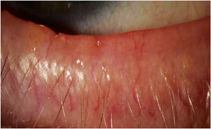
  • Staph Electron MicrographBacterial Blepharitis – This type of blepharitis occurs from a either an acute or a chronic bacterial infection of eyelid margin at the base of the eyelashes.  Typically this infection comes from gram-positive Staphylococcal or simply “Staph” bacterial with the most common culprit in this family being Staph Aureus (electron micrograph image right).  These Staph bacterial are extremely common on the human skin and in the eyes, but inflammation or infection comes from over population or hyper sensitivity to these bacterium.  In acute cases, over population of bacterial causes a sudden, active, and contagious infection of the eyelids with redness, moderate to severe crusting and matting.  This infection can also cause a concurrent infection on the white of the eye called conjunctivitis (“pink eye”) and in severe cases a keratitis which is an infection on the cornea.  Ulcerative BlepharitisAlthough acute bacterial blepharitis is common, since this bacterial is so commonly found on the skin surface it is more common to see a chronic long term hypersensitivity to the offending bacterium.  Chronic bacterial blepharitis causes less overt symptoms but far worse, long term, and irreversible complications including crusting, thickening, ulceration, scarring of the eyelids, and permanent loss of lashes (seen at left).
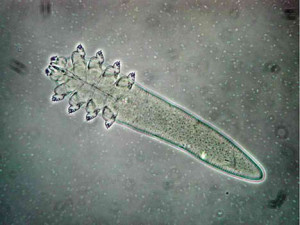
 

  • DemodexDemodex Blepharitis – I promised you parasites!  Like Staphylococcal bacteria, Demodex (pictured at right) are also a ubiquitous and largely benign resident of the human skin.  Demodex are a parasitic mite that spends its life living inside the hair follicles of ALL mammals.   You are probably familiar with mange in dogs?  Yep, that’s demodex (infected follicle at right).  The prevalence of eyelash demodex  in humans increases with age – from 85% prevalence by age 60 to nearly 100% of humans by age 70, but it is commonly found in patients of all age ranges.  Luckily for us, the Demodexvast majority of the time, this parasitic relationship occurs without any problems or symptoms.
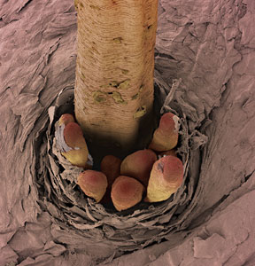
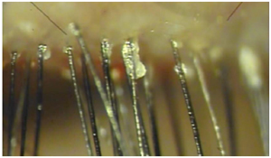
    Problems with this type of blepharitis occur if there is an over population of these mites or if an individual is overly sensitive to the presence of these mites.  The hallmark of Demodex infestation is clear, glistening, cylindrical dandruff at the base of the eyelashes as seen under an eye doctor’s microscope (below).  The waste byproducts and bacteria associated with these mites are what cause the inflammation of the eyelids and leads to discomfort and dryness on the eye.
    There is also a known link between Rosacea and increased overpopulation of Demodex.  The good news is that the life span of a Demodex mite is short (about 20 days); however the bad news is that these mites will mate and reproduce if the infection is not controlled.  Treatment for a mite problem is to cull their ranks with readily available eyelid cleaners containing tea tree oil whDemodex on Lidsich are very effective at lowing the overall population.  It is very common, however, for the symptoms to return as the mites multiply over time.  Therefore, a daily eyelid cleaning
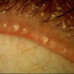
    regimen is very important to keep the mites at bay in the long term.


Thank you for joining us on this trip through the ins and outs of Blepharitis.  We hope we have shed some light on a common condition that can result in some very uncomfortable eyes.  Next week on Eye to the Future, we will take a look at a far more familiar (but not well understood) condition:  “Pink Eye”.

Blepharitis – Posterior Blepharitis

As discussed in our previous post.  Blepharitis has two types:  Anterior Blepharitis and Posterior Blepharitis.

Today we will be discussing the posterior form.

Posterior Blepharitis

Meibomian Gland DiseaseThis condition is also known as Meibomian Gland Dysfunction or simply MGD (no, not the frosty adult beverage).  This form of lid inflammation occurs at the inner edge of the eyelid next to the eyeball where the meibomian (or oil producing) glands are located.  As previously mentioned, these oil glands produce  a very important oil that coats over the watery layer of the tears with every blink.  Without proper amounts or quality of this oil the tear film becomes unstable and evaporates too quickly to keep the eye properly wetted.  As a result, Posterior Blepharitis is the leading underlying cause of evaporative Dry Eye Syndrome (DES).   Inflammation on this part of the eyelid results in these important oil glands clogging and become “capped” or “plugged up” as is shown above.  As these oil glands get clogged, the clear oil they typically produce turns to very thick almost cheBlepharitis - Frothy tearsesy oil (shown above) along with foamy or frothy tears (right).  In addition to problems with the oily layer, the natural bacterial in our tears also tends to get trapped in the clogged glands and can cause an infection of that gland; this is how stys are formed.
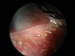
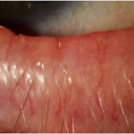
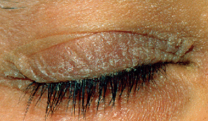
As this chronic inflammatory condition continues over months to years, the lids themselves continue to become more thickened and red, along with prominent engorged blood vessels and eventually the oil glands become dysfunctional all together which results in permanent scarring of the lids (photo below).  Patients with Posterior Blepharitis or MGD are generally older with a long history of ocular symptoms and dry eyes.  Another important note is that this condition is HIGHLY associated with Rosacea since the oil glands on the face where Rosacea occurs are very similar to the oil glands on the eyelids.

Ulcerative BlepharitisClearly, if left unchecked, Posterior Blepharitis can cause substantial damage to the lids and the ocular surface over time.  How do we combat this process?  The answer here isn’t quite so simple.  This is a chronic condition and therefore, there isn’t a single treatment that works best for all patients nor is there a definite “cure”.  Depending on severity, there are a few treatments that are useful in most cases.  This three prong approach includes artificial tears in conjunction with proper eye lid hygiene maintenance and Omega 3 fatty acids (fish oils).  For the artificial tears, it is important to improve the oily layer, therefore we recommended an emollient or “oil rich” tear like Systane Balance or Soothe XP.  We have all been told since childhood to keep soap away from our eyes (and for good reason!  Ouch!), however this fact in combination with the bony orbit preventing access, make the lids the only place on the human body that never gets cleaned.  For our Blepharitis patients, we strongly recommend warm compresses to help loosen the clogged oil as well as routine eyelid cleaning with a dedicated (eye friendly!) cleaner like Ocusoft or Sterlid products.  Omega 3 fish oils are also a natural anti-inflammatory product which will also help the lids to produce better oil, and have the added benefit of being great for heart health.  As is often the case with this condition, if symptoms are more pronounced or if there is a risk of lid scarring or permanent damage, it is often necessary to go further with treatment including prescription eye drops or even oral antibiotics for several months to quell the inflammation and improve the functionality of these glands.

It’s important to remember that there is no “cure” for Posterior Blepharitis.  Sufferers from this condition need routine monitoring and sometimes aggressive treatment to maintain a comfortable and safe eye.

Stay tuned on Eye to the Future for the final installment: Anterior Blepharitis, next week.

Blepharitis – Never heard of it? We think you should!

What if we told you that there is a condition we see in around 40% of patients but was something that you’ve probably never heard about before?  How about if we mentioned that it is the leading cause of evaporative Dry Eye and a main cause of conjunctivitis (“pink eye”)?  Maybe we could peak your interest by saying Blepharitis is more common than Glaucoma, Macular Degeneration, and Retinal Detachments… combined!

Now that you’ve met the contributors to Eye to the Future, let’s kick off this educational blog by turning our eye to this largely unknown, nearly ubiquitous, and often overlooked and under treated condition with an in-depth look at Blepharitis.


What is Blepharitis?

Blepharitis (pronounced “blef-ah-RYE-tis”) is a non-specific term for inflammation of the eye lids and is incredibly common; affecting more than 80 million Americans.  The inflammation and irritation of the eyelids can occur secondary to several different sources which we will discuss in some detail, but regardless of the cause, the inflammation of those lids vastly affects the quality of the tears and often results in redness, ocular discomfort, Dry Eye Syndrome (DES), acute conjunctivitis (“pink eye”), and an overall poor cosmetic appearance of the eye.  The reason for all this is that the eyelids do far more than just blink to protect the eye.  With each and every blink, around 40 meibomian glands (or oil producing glands) on each of the four eye lids produce and excrete an oily substance.  This secretion acts as an “oil slick” that covers over the watery layer of the tears and decreases the rate at which the tears evaporate into the air.  The inflammation and irritation to these eyelids results in the improper function of these oil glands.  Compounding matters, we have all been told since childhood to “never get soap in our eyes” which makes the eyelids the only part of the human body that never gets cleaned.  This results in inflammatory compounds, oils, and overall crusting building up resulting in further inflammation the surface of the eye and more symptoms.  This vicious cycle leads to eyes in bad need of some TLC…

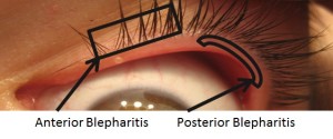
Types of BlephAnterior and Posterior Blepharitisaritis

There are two main classifications of Blepharitis- Anterior and Posterior Blepharitis – and these terms are simply based on the location of where the lid inflammation is occurring.  Anterior Blepharitis occurs at the front outside edge of the eyelid at the base of the eye lashes and/or further up onto the sensitive eye lid skin.  Posterior Blepharitis by contrast occurs at the inner edge of the eyelid next to the eyeball where the meibomian (or oil producing) glands are located.

Now that we know what Blepharitis is and where it occurs, in the next two installments of this series on Blepharitis we will delve further into the different types of blepharitis, what causes each type, what symptoms to watch out for, how these conditions are treated… and maybe even dabble into a little discussion on eyelid parasites!  YIKES!

Stay tuned as we keep an Eye to the Future.


Meet Dr. Roberts

Dr. Keith Roberts

Dr. Keith Roberts is a Louisville-area native, born and raised in Clarksville, Indiana. Upon graduation from Clarksville High School, Dr. Roberts attended Indiana University Southeast earning a B.S. in biology. After completing undergraduate studies in 2004, Dr. Roberts attended the Indiana University School of Optometry, receiving a Doctor of Optometry degree in 2008.

During his tenure at the Indiana University School of Optometry, Dr. Roberts spent time training at Eye Surgeons of Indiana, a tertiary care referral center in Indianapolis, focusing on treatment and management of ocular disease, as well as pre and post-operative care for cataract and Lasik patients.

In addition, Dr. Roberts received training in primary eye care at the Indianapolis Eye Care Center in Indianapolis and the Ireland Army Hospital in Fort Knox, KY. Clinically, Dr. Roberts has a special interest in the treatment and management of glaucoma, as well as non-surgical emergency eye care.

While he is out of the office, Dr. Roberts enjoys spending time with his wife and their two little girls. In addition, Dr. Roberts is an avid sports fan and enjoys fishing when time permits.


Meet Chris

Chris SowderChris Sowder, our office administrator, brings to our practice nearly 20 years of experience. Chris has an Associate of Science degree from Indiana School of Optometry’s optician/technician program. Following school she was an assistant retail manager at a large optical chain, providing her with customer service and optician/dispensing experience. Chris then made a career move to the medical side of optometry as a lead ophthalmic technician for Bennett and Bloom Eye Centers. This experience taught her the importance of early treatment for ocular disease. Most recently, Chris has worked for a busy private practice where she was cross-trained in every department. It has always been Chris’s goal to maximize her college degree and work as an office manager of a locally owned, state-of-the-art Optometry practice. The road she has taken to get here gives her every advantage helping her patients get the most out of their eye care needs. This born and raised Hoosier spent a few years in Bloomington and Indianapolis, but is now proud to call her hometown of Pekin, Indiana home once again. Chris is married and has three school-aged children to keep her busy in her time away from work.

Meet Dr. Wolf

Dr. Nick Wolf

Dr. Nicholas Wolf is a native Hoosier who completed his undergraduate degree in Biochemistry from Indiana University Bloomington and then went on to attend the Indiana University School of Optometry, where he graduated with a Doctorate of Optometry in 2008. He began practicing in Indianapolis before moving to Floyd County in 2012.

During his training at Indiana University, Dr. Wolf received extensive training in eye disease diagnosis and management at Eye Surgeons of Indiana based in Indianapolis as well as at the Dayton, Ohio VA eye clinic.

Dr. Wolf practices full scope optometric care with a special interest in ocular disease evaluation and treatment, including glaucoma, macular degeneration, as well as cataract and laser refractive surgery co-management. Dr. Wolf is also very comfortable and practiced with pediatric populations as well as specialty contact lens fitting. He has continually upgraded his techniques and knowledge in these arenas, as the profession of optometry has expanded, to provide his patients the most advanced possible care.

Dr. Wolf prides himself on his professional, personal, and thorough examinations. He strives to make patients feel welcome and comfortable while he addresses their eye care needs and educates them about their visual system.

Dr. Wolf lives in Floyd county with his wife Edie and is an avid sports fan and counts himself as a loyal Indianapolis Colts and Hoosier faithful. As time permits, he is also an avid woodworker and many of the pieces seen in the office came from his home workshop.