Precision Family Eye Care – Ribbon Cutting

On Friday, July 11, 2014, Precision Family Eye Care had its Ribbon Cutting and is now officially open for business!!

photo 1                         photo 1 (1)

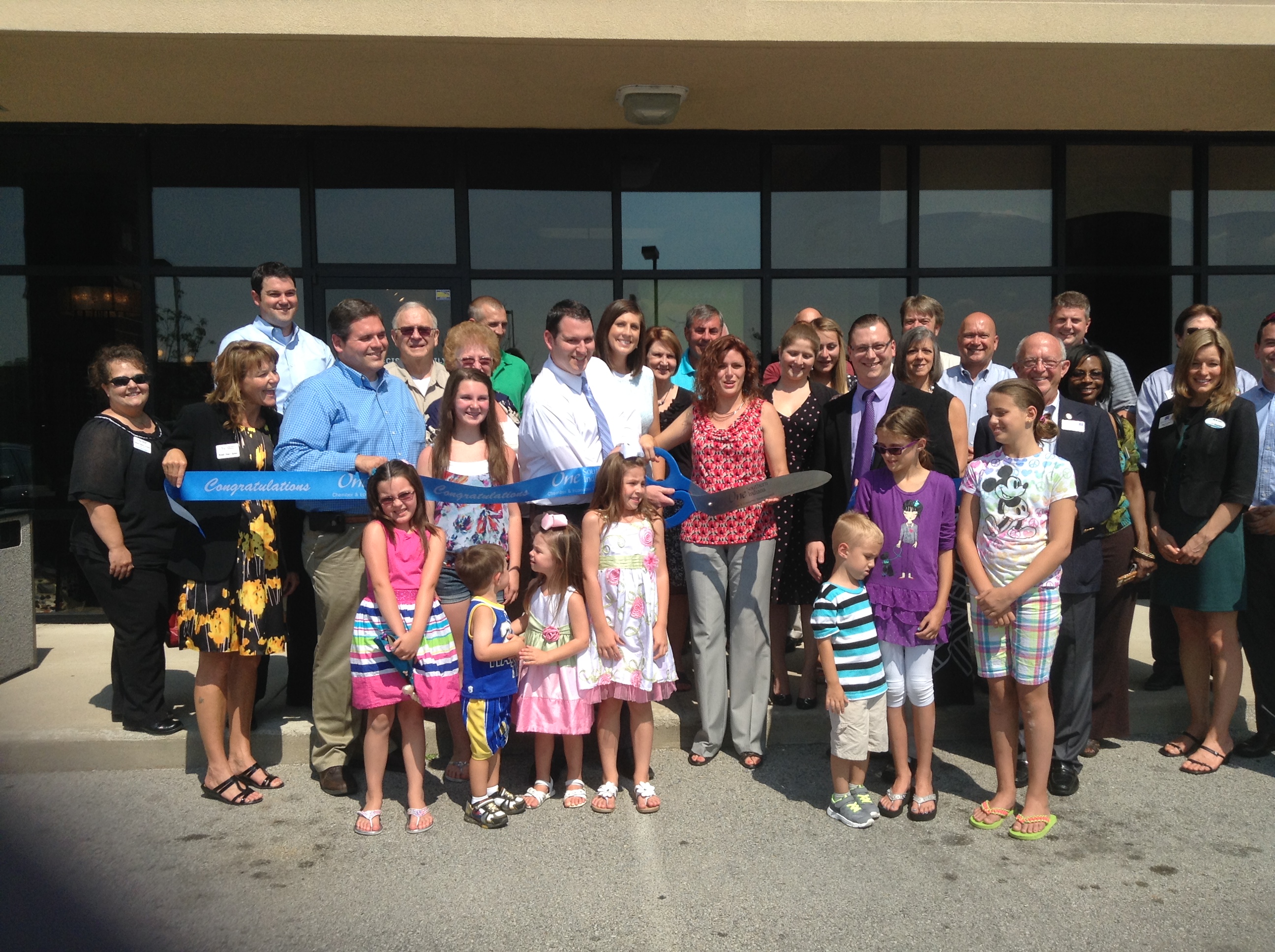
Special thanks to Senator Ron Grooms and Representative Ed Clere for taking time out of their extremely busy schedules to celebrate with us.  We were also joined by Wendy and the rest of the terrific staff from the One Southern Indiana Chamber of Commerce who did a fantastic job of organizing and structuring the event.   Thanks as well to all the 1SI ambassadors and fellow professionals and business owners who graciously spent their Friday afternoon with us and offered us their support and congratulations.  Most importantly, we would like to thank our families for encouraging us, we wouldn’t have gotten this far without your support.

At Precision Family Eye Care, our mission statement is to provide clinical excellence, quality products and unmatched personal and professional services to give our patients the best opportunity for a lifetime of healthy eyes and excellent vision.  We call this area home and look forward to providing comfortable, personalized, and comprehensive care to the wonderful people of this community.


Nevus


Nevus

Statistics and Associations

  • Relatively low risk of melanoma; approximately 4 in 1000
  • Suspicious lesions include growth (after puberty), significant elevation, underlying fluid, overlying orange pigment, close proximity to optic nerve and resultant visual symptoms

Management

  • Photography for baseline
  • Suspicious Nevi should be followed with serial photography to detect any changes

Retinal Detachment


Retinal-Detachment
Statistics and Associations

  • Separation of retina from underlying tissues
  • Risk factors: lattice, nearsightedness, PVD, vitreous hemorrhage, retinal breaks (holes/tears), trauma
  • 30% association with lattice degeneration

Management

  • Surgical intervention
  • If macula is attached, emergent treatment is indicated (within 24 hours)
  • If macula is detached, urgent treatment is indicated (within 48 hours)

Lattice Degeneration


Lattice-Degeneration
Statistics and Associations

  • Thinning of peripheral retinal due to traction
  • Present in 10% of population
  • 25% develop holes in retina
  • More common in nearsighted people
  • 40% of retinal detachment patients also have lattice degeneration

Management

  • Monitor as indicated
  • Patient education regarding signs/symptoms of retinal detachment

Optic Atrophy


Optic-Atrophy
Statistics and Associations

  • Can be: traumatic, compressive (intraorbital/intracranial mass), hereditary (bilateral), toxic (bilateral), nutritional (bilateral), or secondary to various systemic situations/diseases
  • Vision variable (20/20 – 20/400)

Management

  • Consider neurology consultation

AION (Anterior Ischemic Optic Neuropathy)


AION-Anterior-Ischemic-Optic-Neuropathy
Statistics and Associations

  • Two forms: arteritic (emergency) and non-arteritic
  • Non-arteritic associated with high blood pressure (40%) and diabetes (20%)
  • Occurs in people older than 55 (older than 60 for arteritic)
  • Fellow eye involved in 25%-40% (non-arteritic)
  • Vision loss usually permanent

Management

  • Visual Field testing
  • Consider STAT lab testing (ESR, CRP, CBC), check blood pressure
  • Consider daily aspirin for non-arteritic AION

Optic Nerve Head Drusen


Optic Nerve Head Drusen
Statistics and Associations

  • Buried material within the substance of the optic nerve
  • 50% develop visual field defects
  • Visual field defects can mimic glaucomatous defects
  • Defects may change as the substance moves forward

Management

  • No treatment necessary
  • Monitor with routine visual fields to rule out glaucoma

Papilledema


Papilledema
Statistics and Associations

  • Bilateral swelling of the optic nerve due to increased intracranial pressure
  • Commonly associated with headaches, TVO’s, and pulsatile tinnitus
  • Typically caused by an intracranial mass, malignant hypertension, or idiopathic intracranial hypertension

Management

  • Visual field (enlarged blind spot), Optic nerve imaging (OCT)
  • Emergent MRI of brain and orbit, lumbar puncture
  • Check blood pressure

Toxoplasmosis


Toxoplasmosis
Statistics and Associations

  • Necrotizing inflammation of the retina caused by a parasite
  • Can be acquired (poorly cooked meat) or congenital

Management

  • Amsler grid for daily monitoring
  • No treatment necessary if inactive
  • Consider referral to retinal specialist if active

Histoplasmosis (POHS)


Histoplasmosis-POHS
Statistics and Associations

  • Scarring in the retina secondary to a previous fungal infection
  • Common in individuals raised on a farm (around chickens or other birds)
  • Endemic in Ohio and Mississippi river valleys
  • Fungus lies dormant with the potential to reactivate later
  • Possibility of new blood vessel growth under the retina at scar location in macula.

Management

  • No treatment necessary if inactive with no new blood vessel growth
  • Refer new blood vessel growth for treatment with anti-VEGF injection