Idiopathic Central Serous Chorioretinopathy (ICSC)


Idiopathic-Central-Serous-Chorioretinopathy-ICSC
Statistics and Associations

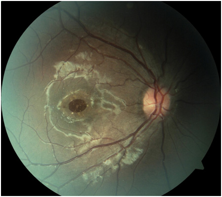
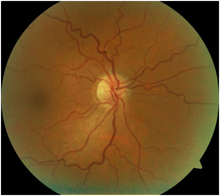
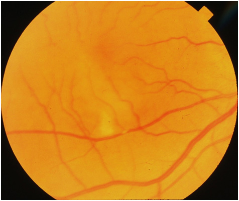
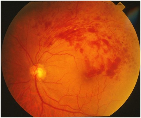
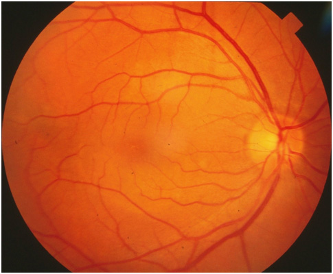
  • Leakage of fluid leading to a blister in the subretinal space
  • Commonly associated with type A personality and recent stress
  • Most common in males (10:1), more common in whites (rare in blacks)
  • 45% recurrence rate within one year
  • 5% develop new blood vessel growth under the retina (SRNVM)
  • 95% end up with vision better than 20/40

Management

  • Amsler grid for daily monitoring
  • No treatment necessary (usually resolves within 3 months)

Macular Hole


Macular-Hole
Statistics and Associations

  • Risk factors: macular swelling, macular traction, trauma, idiopathic
  • 10-20% chance of developing macular hole in fellow eye

Management

  • Amsler grid for daily monitoring
  • No treatment for mild macular holes due to the possibility of spontaneous closure
  • Surgical intervention (PPV with membrane peel, air-fluid exchange, gas injection). Requires face down positioning for weeks
  • Poor prognosis if macular hole has been present for one year

Hypertensive Retinopathy


Hypertensive-Retinopathy
Statistics and Associations

  • Common with blood pressure above 140/90
  • Generally asymptomatic
  • Sclerosed arteries, vessel tortuosity, and vessel crossing abnormalities are the most common changes
  • Optic nerve head swelling, dilated tortuous veins seen with malignant hypertension

Management

  • Medical consult to treat underlying hypertension

Branch Retinal Artery Occlusion


Branch-Retinal-Artery-Occlusion
Statistics and Associations

  • Associated with high blood pressure (67%), Carotid Occlusive Disease (25%), Diabetes (33%), Cardiac Valve Disease (25%)
  • 80% will end up with vision better than 20/40 if macular area not involved
  • Most will have permanent visual field defects
  • Mild association with stroke

Management

  • Medical Evaluation with complete cardiovascular evaluation including electrocardiogram, echocardiogram, carotid doppler studies

Branch Retinal Vein Occlusion


Branch-Retinal-Vein-Occlusion
Statistics and Associations

  • Obstruction of the retinal venous drainage system usually due to a hardened artery pressing on a retinal vein
  • Associated high blood pressure (60%), Glaucoma (40%), Diabetes, Heart Disease, rarely Hypercoagulable States (conditions that cause blood to be thicker than normal)
  • 50% will have Macular edema, 20% will develop epiretinal membranes
  • 50% will have better than 20/40 visual acuity

Management

  • Medical evaluation with attention to cardiovascular system to treat underlying cause
  • If macular involvement, intraocular injection of anti-VEGF agent.
  • New blood vessel growth may require laser treatment

Central Retinal Vein Occlusion


Central-Retinal-Vein-Occlusion
Statistics and Associations

  • Obstruction of the retinal venous drainage system usually due to a hardened artery pressing on the central retinal vein
  • Associated high blood pressure (60%), Glaucoma (40%), Diabetes, Heart Disease, rarely Hypercoagulable States (conditions that cause blood to be thicker than normal)
  • Major complications include new blood vessel growth and macular swelling

Management

  • Medical evaluation with attention to cardiovascular system to treat underlying cause
  • Ocular injection of anti-VEGF agent now standard of care
  • Gonioscopy at each visit
  • New blood vessel growth may require laser treatment

Posterior Vitreous Detachment (PVD)


Posterior-Vitreous-Detachment-PVD
Statistics and Associations

  • Detachment of the vitreous from the retina
  • 53% by age 50, 65% by age 65
  • 10% – 15% risk of retinal break with acute PVD
  • 70% risk of retinal break if vitreous hemorrhage is present

Management

  • For acute PVD: follow in 2-4 weeks to rule out retinal break
  • Patient education regarding signs/symptoms of retinal detachment

Asteroid Hyalosis


Asteroid-Hyalosis
Statistics and Associations

  • Calcium phosphate material attached to vitreous
  • 75% unilateral
  • 30% association with diabetes
  • Typically no effect on vision

Management

  • No treatment necessary

Pseudoexoliation Syndrome


PseudoexoliationS-yndrome
Statistics and Associations

  • Characterized by an abnormal accumulation of white fibrillar material in the anterior chamber
  • Commonly seen after fifth to sixth decade of life
  • Increased intraocular pressure changes noted in 50% of pseudoexfoliation syndrome patients within ten years of diagnosis

Management

  • No treatment recommended unless glaucomatous changes occur
  • Laser treatments quite effective as well as topical medications

Pigment Dispersion Syndrome


Pigment-Dispersion-Syndrome
Statistics and Associations

  • Liberation of iris pigment cells into anterior chamber can clog drainage canal and increase pressure
  • Typically first seen in white males between 20–40 years old
  • Visual fluctuations common with physical exertion
  • Pigmentary glaucoma occurs in approximately 10-20% of patients
  • If glaucomatous changes occur from PDS, usually happen before fifth decade of life

Management

  • No treatment recommended unless glaucomatous changes occur
  • Laser treatments quite effective as well as topical medications