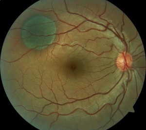
Solar Eclipse Eye Safety!!


In about a month, the Kentuckiana region is directly in the path of The Great American Solar Eclipse!!


solar eclipse


This solar eclipse will occur in our area on August 21st at 12:21 PM EST.


This eclipse will cross the entire continental US.  While the eclipse will be visible from nearly every state, it will be especially spectacular in the Louisville area.

Here there is predicted to be a 90-95% partial eclipse, and if you head to southwestern Kentucky (around Hopkinsville) it will be a complete solar eclipse for about 2 minutes!

These events area extremely rare with the last total eclipse occurring here back in February 26th, 1979!!

eclipse map


While this once in a life time event is amazing, it is also EXTREMELY DANGEROUS!

It is absolutely critical that this miracle of nature be viewed through an approved device.


These devices include:

  1. ISO Certified eclipse glasses (certification 12312-2:2015)
  2. Welders Shield or Filter with shade #12-14
  3. Viewed indirectly with a Pinhole Camera – A great project for kids!  (NASA project link)

Wearing sunglasses or even ‘taking a quick peek’ can cause irreparable damage.

Don’t Risk It!!


Solar Retinopathy
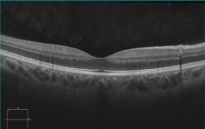

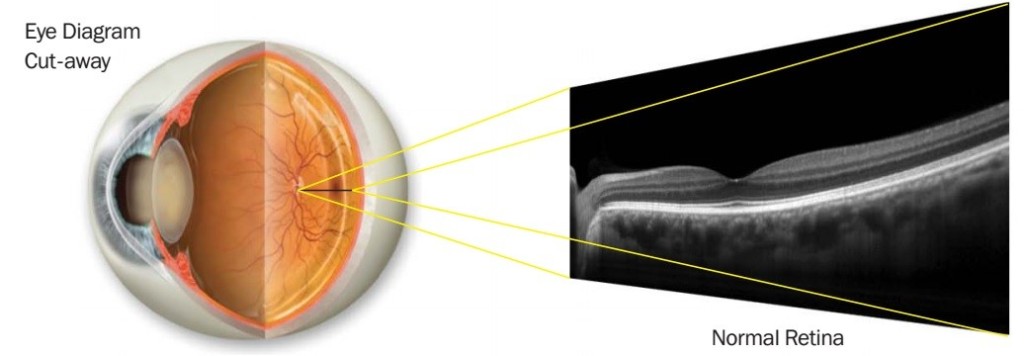
normal oct


The eye focus light that enters the eye back onto the retina.  In particular, the light is focused most strongly onto the macula, which is the center part of the vision (normal imaging of macula above).
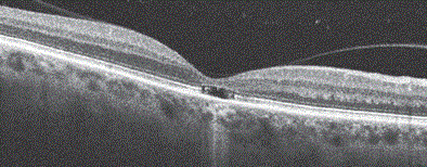


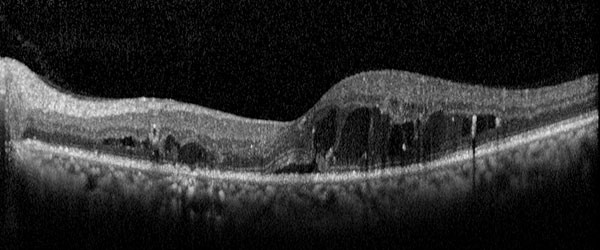
However, when high intensity, direct sunlight is focused on the macula, it heats up the pigmented layer underneath the retina.  This layer gets so hot from the high intensity light that it actually burns the cells that sense light in the center part of the vision (see above).

When this area is damaged, vision will be decreased and you may noticed a blind spot in the center of your vision.  Sometimes this condition improves over time but often the vision does not completely return and permanent vision loss occurs.


If you would like to know if the eclipse glasses you have are safe, please email or stop by our office and we’d be happy to talk with you further.


Be Safe and Enjoy the Eclipse!!

Dr. Wolf


Scleral Contact Lens

Scleral Contact Lenses to the Rescue

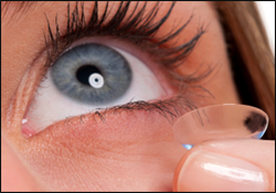
Contact Lens FittingToday on Eye to the Future we will be discussing a type of contact lens you probably haven’t heard of before, but one that has helped many of our patients live fuller, happier, and more visually productive lives – Scleral Contact Lens.
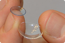
There are an estimated 38 Million contact lens wearers in the United States.  For most of those, contact lenses are seen as a cosmetic privilege, allowing freedom from the need for glasses, to look better in pictures, or even to change their eye color!


For many though, hard contact lenses are a necessity to function and live a normal life.


Hard Contact Lenses are Still Around?!?

Most contact lenses wearers think hard contact lenses are a thing of the past.   With the vast improvements in soft contact lens technology, that is partially true.  However, there are many eye diseases and conditions that require hard lenses.  Soft lenses or even glasses simply DO NOT provide adequate vision or comfort.


Scleral Contact Lenses

Scleral Contact LensesA newer type of hard lens for eye disease is the scleral contact lens.
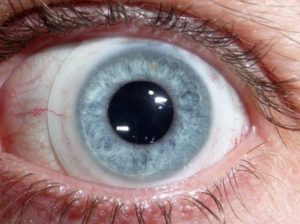
As the name implies, a scleral contact lens is a hard lenses that rest entirely on the white of the eye, called the sclera.  They are much, much larger than traditional hard lenses (left).

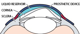
These lenses vault over and do not even the touch the cornea, unlike all other contact lenses.  A saline solution then fills in the area between the scleral contact lens and the cornea.


Scleral contact lenses are most useful for two eye problems:

Keratoconus and Corneal Ectasia

Severe Dry Eye and Sjögren’s syndrome


Corneal Ectasia (Bulging) and Keratoconus

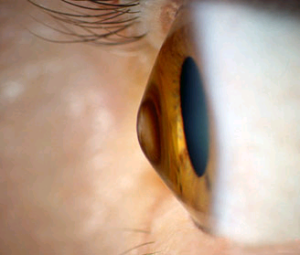
keratoconusThe cornea is the clear front surface of the eye.  Normally, the cornea has a round regular shape, like a ball.  Other times the cornea is more egg or football shaped.  This is called an astigmatism and is also very normal.
Sometimes, however, the structure of the cornea is not strong enough to hold a stable, rounded shape.  The normal pressure inside the eye then causes the cornea to irregularly bulge outward in places like a cone.

This condition is called keratoconus (right) and is the most common cause of ectasia affecting 1 in 2000 Americans.  About 50% of the time genetics is involved.  In reality, it is most closely linked to eye rubbing, especially as a child.

In addition to keratoconus, corneal bulging (ectasia) can also occur after refractive surgeries like LASIK, PRK, and especially after RK or radial keratotomy, as well as after corneal transplants.


Why is Scleral Contact Lens Useful for Keratoconus/Ectasia

PROSEThe cornea is the first surface that light goes through.  The bulging irregular surface from ectasia heavily distorts the image going through it.
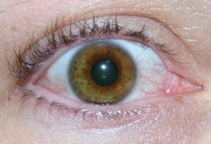
This causes substantial blurring and glare for a patient with ectasia.

Glasses cannot compensate for the irregular surface of the eye.  Soft contact lenses do no better as they simply lay over top and conform to the bulging cornea.

Scleral contact lenses act as a PROSE or Prosthetic Replacement for the Ocular Surface Ecosystem (image above).  A scleral contact lens vaults over the irregular cornea and fluid fills up the space in between, essentially creating a new, prosthetic cornea.  Pretty cool, huh?  And they work GREAT!  Often scleral contact lenses will take an ectasia patient from not passing a drivers test down to the perfect 20/20 line and sometimes better!


Studies show that patients with ectasia who are switched to a scleral contact lens showed an 80% improvement in comfort and vision and a 90% improvement in overall satisfaction and quality of life.


Scleral Contact Lenses (Scleral Shell) for Severe Dry Eye and Sjögren’s Syndrome

dry eyeDry eye syndrome is the most common cause of eye discomfort, affecting over 25 million Americans and 1 in 4 people over 60.

Mild to moderate dry eye is often successfully treated with artificial tears, prescription medications (like Restasis and steroids) or through simple in office procedures.

However, with severe dry eye, traditional options are often not adequate.  In these cases, a scleral shell is used, especially for patients with Sjögren’s syndrome.


Sjögren’s Syndrome

Sjögren’s syndrome is an autoimmune disease where the body attacks the water secreting glands of the body.  Most often affected are the salivary glands of the mouth and the lacrimal gland that produces the “water” part of the tears.

Because of the continual inflammation in these glands, Sjögren’s syndrome patients often have severe dry mouth and dental problems as well as incredibility dry eyes where traditional dry eye therapy is simply not effective enough.


Scleral Contact Lens (Scleral Shell) for Severe Dry Eye

scleral contact lensPatients with severe dry eye require copious amounts of lubrication to achieve comfort and provide sustained, acceptable vision.

A scleral contact lens vaults over the sensitive cornea.  The highly wettable surface of the lens provides clearer vision without the visual fluctuations often experienced with severe dry eye.

Most importantly, the lenses traps lubricating saline under the lens and against the sensitive cornea.  This serves as a soothing water bath, with constant lubrication for the front surface of the eye.


If you or someone you know suffers from keratoconus, corneal ectasia, severe dry eye, or simply cannot obtain adequate vision with glasses or traditional contact lenses –

scleral contact lens may offer a solution.


For additional information or to schedule a fitting for scleral contact lenses, please contact us.

[email protected]

As always, thank for tuning into Eye to the Future and I’ll see you on the next installment.

  •  Nick Wolf, OD

More information about Scleral Contact Lenses

Scleral Lens Education Society

All about Vision – Keratoconus

Our webpage on Scleral Lenses with additional links


The Mantis Shrimp


The Amazing Eyes of the Mantis Shrimp


peacock mantis shrimpThink you see pretty well?

At Precision Family Eye Care we strive to make all of our patients see their BEST with glasses and contact lenses.

Unfortunately the human eyes can’t even hold a candle to the insane visual capacity of the Mantis Shrimp.

Today on Eye to the Future, we will explore the MOST ADVANCED EYES on the PLANET!!  Meet the Mantis Shrimp!!

The Mantis Shrimp’s name is really a misnomer as it is neither a Mantis, nor a Shrimp, but is actually a close relative of the lobster.  However, it takes one look to see this name is a pretty apt description for this amazing creature.

The Peacock Mantis Shrimp is pictured above. They will typically grow up to 8 inches long.  I’ve personally had two of these as pets, they’re characters…grumpy characters.


What’s So Special?


mantis shrimpThe mantis shrimp has two eyes, same as us, but that is where the similarities end.

First, the eye stalk is directional and capable of rotating each eye independently up to 70 degrees in any direction.  This allows each eye the ability to view its entire surroundings, kind of like the periscope on a submarine.

Humans, and mammals in general, have two eyes with one pupil each to focus light.  Put two eyes together and you have binocular vision which allows us to sense depth in our three dimensional world.

However, EACH eye of the mantis shrimp has THREE pupils (above)!  This means that each of the mantis shrimp’s eyes is capable of Trinocular vision and thus sensing depth with each eye simultaneously.  Focus both eyes on the same thing and you have Hexnocular vision!


Thermonuclear Bomb of Color


As you can see from the photo above and below, each eye of the mantis shrimp is separated into three zones.

What’s going on with that?

Here is where the eyes of the mantis shrimp get REALLY interesting.

The human eye has three types of photoreceptors (cells that sense light) for seeing color.  We have Red, Green, and Blue.  The combination of signals from these three cell types allow us to see all the colors in the visible color spectrum.  Dogs have one less than us with two (no, dogs aren’t actually color blind) and butterflies actually have 5.  To our three, the mantis shrimp has 16 different photoreceptors!!  As one researcher put it, they see a Thermonuclear Bomb of Color!  Here’s how…

mantis shrimp eyes1The mantis shrimp’s eye is separated into three different sections.  The upper and lower hemispheres are used mostly for sensing motion and forms.  The central area is called the midband and contains 6 rows of specialized clumps of photoreceptors.
The first 4 rows contain the color sensing photoreceptors that allow mantis shrimp to see colors in hues we couldn’t imagine and even allows it to see near infrared and ultraviolet (UV) light.  These rows also contain filters that the mantis shrimp can use to adjust their visual input – like putting on different pairs of colored sunglasses.  And if that was not amazing enough, row 5 and 6 of the midband even allow the mantis shrimp to see various polarized light!


How does this all work together?


Say you have a mantis shrimp, it’s crawling around the ocean floor doing little mantis shrimp stuff.

The upper or lower areas of the Mantis Shrimp’s eyes detect movement, like, say, a potential meal.  The mobile eye stalk then allows the shrimp to move the eye to use the midband, scanning the target with it’s incredible array of photoreceptors to glean additional information… AND, each eye can do this independently.

Here is a terrific video of a mantis shrimp scanning it’s surroundings to illustrate how this works!

In fact, the brain of the mantis shrimp is actually very, very small by comparison.  Their eyes contain more nervous tissue and it’s thought may actually “control” things.  Cool stuff.

So there you have it, the most advanced eyes on the planet…  But, wait!  There’s more!!


Mike Tyson of the Sea


mantis shrimp saddleSuperlatives don’t stop there with these little critters!
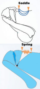
Mantis shrimp are also the Guinness world record holders for the FASTEST attack in the animal kingdom.  The mantis shrimp stores energy in highly specialized cartilage called the saddle at the base of the claw (right).  When this pent up potential energy is released, the strike occurs with the same speed  as a 22 caliber bullet!

They use this attack to break open clams, mussels, crabs, stun/kill fish, or just to take a whack at whatever looks at them funny.

Their strike is so fast that water literally boils as it moves out of the way.  This causes a secondary shock wave that causes more damage to the prey and even creates light and heat.

This attack occurs faster than the human eye can detect it.

Here is a video with a very high speed camera showing the attack as well as the cavitation bubbles and light produced by the strike.. Amazing.

Their attacks are known to break aquarium glass…in addition to anything else that crosses it’s path.  In the aquarium, fishing, and scuba diving world, mantis shrimp are commonly referred to as “Thumb Splitters.”

They are armed to the teeth and aren’t afraid to break a finger if you come to close!!


I hope you enjoyed this slightly off topic discussion.  When it comes to animals, Mantis Shrimp are not particularly cute and are definitely not cuddly, but they are an apex predator with some incredibly specialized killing equipment.

Until next time!

Nick Wolf, OD

[email protected]

– AMAZING photography of the Mantis Shrimp credited to Michael Bok.  Here’s is his website.  Videos embedded where taken from Youtube.

How to stop myopia progression?

appointment-300x150(1)


“Why do my child’s eyes keep getting worse every year and is there a way to stop it?”  


It’s a question we are often asked and it’s a perfectly reasonable concern…

Nearsightedness or myopia progression is on the rise in the US.  Studies from the 1970’s showed that 25% of the country was nearsighted but by 2010 that number had ballooned up to 42%!!  That’s an increase of 66%!!  Additionally, the total amounts of nearsightedness had also dramatically increased!

Considering the effect of high myopia on learning, quality of vision, as well as a substantially increased risk of Glaucoma and Retinal Detachments – it seems logical to consider how to stop it.

Today on Eye to the Future, we will look at Myopia.  What is actually happening?  What causes it?  And what can we do to slow myopia progression?


What is Actually Happening in Myopia?

Nearsightedness or Myopia is simply put, long eyeballs.  It’s really nothing more exotic than that.

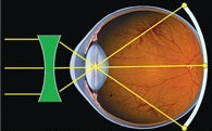
In nearsighted individuals, the eye grows axially or front-to-back.  This growth primarily occurs because of something called peripheral defocus.

peripheral defocusSince the eye grows front-to-back, the whole eye isn’t growing, just the parts around the outside of the eye in the peripheral retina.  If light entering the eye doesn’t fall properly on this part of the retina (shown left), it stimulates the eye to grow backwards to focus the light.  Unfortunately, this growth causes the light falling in the center part of vision to be out of focus resulting in fuzzy distance vision.
Not surprisingly, eyes tend to grow most as the rest of the body does, leading to faster nearsighted progression during childhood, adolescence, and puberty.


What Causes the Eye to Grow?

Now there is the million dollar question.  There really isn’t a single cause.  Genetics play a very large role, but the bigger thought is that the increase in near tasks  may be the main driver.

The reason is that when we focus our eyes to see things up close, the amount of peripheral defocus explained above increases causing a greater likelihood of myopia developing.  This would certainly explain why nearsightedness has increased so strongly in the past several decades as the youth have given up playing outside for computers, iPads, and cell phones.

Now parents, before you confiscate Timmy’s DS, it’s important to note that peripheral defocus increases with ALL up close tasks.  A 2010 survey study out of China looked at myopia development with education level and showed that myopia was present in 32% of elementary students but increased in over 80% of college graduates!!  Books may make you smart but they may also make you nearsighted…


What Can be Done to Stop Myopia Progression?

The answer is simple, stop peripheral defocus, the implementation of that however, is more difficult.  In the past many things have been tried.

Making kids go play outside more, wearing hard contact lenses, under correcting children in glasses, even giving kids bifocals – all these have largely been discredited or are difficult to study effectively.

So what does work?

Atropine

The best proven way of stopping myopia progression, is with atropine.  Atropine is a very strong dilating drop that temporary paralyzes the eyes focusing muscle which stops peripheral defocus.

Strong atropine has been shown in studies to cut down on the progression of myopia by 81%!!

While this certainly works, staying dilated throughout the entirety of your childhood is… less than ideal.  Recently, researchers have been experimenting with heavily diluted atropine which works almost as well.

 Pretreat Topo

Treated Topo


Orthokeratology

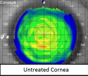
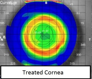
A newer advancement in the fight against myopia progression is Orthokeratology (Ortho-K).  This method uses large, hard contact lenses that children wear at night to reshape the surface of the eye so no glasses or contacts are needed throughout the day.  Think of it like a retainer for your eyes or even temporary LASIK.

One of the surprise benefits of Ortho-K is that this technique drastically reduces the amount of peripheral defocus.  Because only some of the cornea is molded, light is focused more evenly on the inside of the eye (seen above).

In controlled studies, Ortho-K reduces the progression of myopia by 50%!

The downside of Ortho-K is that it is very expensive, vision often fluctuates as the cornea “molds back” through the day, and children have to wear lenses overnight which increases the risk of infection.

Bifocal Contact Lenses

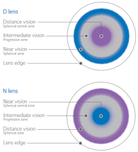
Bifocal Contact LensesThe newest, and most exciting advancement in myopia progression control is through the use of Bifocal Contact Lenses.  Click here for more about bifocal contact lenses.

The short of how this works is that bifocal contact lenses have different focusing rings that allows people going through the curse-of-the-40’s to see up close again.

For kiddos, the ring design, similar to OrthoK, reduces the amount of peripheral defocus and has also been shown to cut nearsightedness progression in half!

The benefits here is that bifocal contact lenses are readily available, inexpensive, worn only during the day, and will correct for distance vision just like any other contact lens!  Cool stuff!

I hope you have enjoyed this detailed explanation of Myopia and what current research says about controlling it’s progression.

If your child is experiencing drastic changes in their myopia from year to year, I highly recommend to speak with your optometrist about ways to slow this awful progression.

If you have any questions or comments, feel free to contact me at [email protected].

Take care of those eyeballs!

  • Nick Wolf, OD


iWellness from Optovue

iwellness1

The three leading causes of blindness in the US are Diabetic Retinopathy, Macular Degeneration, and Glaucoma: all three have NO symptoms initially.

This reason is why eye exams, including a thorough retinal evaluation, are important to protect vision. In an effort to provide a more complete and thorough ocular health examination, Precision Family Eye Care has incorporated the iWellness retinal scan as an integral part.

iWellness

Although the retina is only 3 human hairs thick, this painless three second scan of the eye shows the retina in incredibly high, MRI quality, detail. The iWellness technology can often detect subtle disease changes earlier than microscopes and can even identify the very first changes from Glaucoma twice as early as traditional testing methods.

Because of the benefits of early detection, we have incorporated the iWellness retinal imaging as a NO CHARGE addition to every Complete Ocular Health Exam.

Diabetic Retinopathy and Edema

iWellness Diabetic Retinopathy

Macular Degeneration

iWellness Macular Degeneration


Link for additional information about the iWellness Retinal Scan.


Top Ten Uses for Extra FSA Funds


It is that time of year to check your Flexible Spending Account (FSA) balances!

koali 2December is a hectic time for Americans – shopping, planning, cooking, cleaning, travel plans – but there is one task that so often goes unfinished at this time of year:  Using up your Flexible Spending Account.

Over 14 million Americans participate in a Flexible Spending Account, and for good reason!  With the ever increasing cost of deductibles and non-covered services, these tax free havens certainly do help defray the cost.

Unfortunately these Flexible Spending Accounts do come with one, rather large, catch… you need to spend it, or lose it!

The vast majority of the funds need to be spent by December 31st or they’re gone.  Up to $500 can be used up until March 31st of the next year.  And unfortunatly, A LOT of money is lost…

It is estimated that upwards of $150 – $200 MILLION dollars is lost every year!!

The good news, is that there are some excellent, healthy, productive, and proactive uses for those extra funds.


Top 10 Creative Uses for Extra Flexible Spending Account Funds


1)   Eye Care!  This is an Optometric Blog after all!:  Exams, prescription glasses and sunglasses, contact lenses, dry eye treatments, you name it!

2)  Dental Care:  While you can’t use Flexible Spending Account money for teeth whitening.  A routine cleaning is certainly good, along with dental work you’ve been putting off, or even investing in a new set of dentures.

3)  Try Something Different:  Many Flexible Spending Accounts can be used to go see your chiropractor or even try acupuncture!

4)  Take a try at “Being” the Doctor:  Small, home medical equipment is fair game with Flexible Spending Account funds.  Things like thermometers, stethoscopes, blood pressure monitors, etc.

5)  Trick out that Medicine Cabinet: The vast majority of OTC medications are considered acceptable: cold medicine, contact solutions, eye drops, decongestants, aspirin, antacids, even acne medications!

6)  Stock up on Diabetic Testing Supplys: If you’re a diabetic, here’s the opportunity to get a new blood sugar monitor and stock up on testing strips.

7)   “Honey are we out of band-aids??”:  Everyone needs a proper first aid kit!  Use you extra Flexible Spending Account funds for band-aids, antibacterial cream, gauze, bandages, wraps, etc.  Make several kits and put one in every car.  You never know where you’ll be when you meet need a little triage kit.

8)  LASIK:  Although a leap of faith, Flexible Spending Accounts are a great way to defray the cost of elective ocular surgery.

9)  Ease those aching Joints:  Joint creams, heating pads, hot and cold packs, even eye masks are okay for Flexible Spending Account bucks!

10)  Be Proactive:  Allergy medications, many vitamins, even some sunscreens.  Whatever, you think you’ll need in the coming year.


Thanks for stopping by and we hope you have a Happy, Healthy, and Blessed Holiday and New Year.

– Nick Wolf, OD


Choroidal Nevus aka “Freckle in your Eye”


You’re telling me I have a freckle ‘INSIDE’ my eye?!?


Choroidal Nevus If I had a dollar everytime I heard that from patients… well I sure wouldn’t be rich, but it actually does happen a lot!
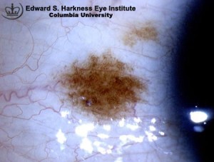
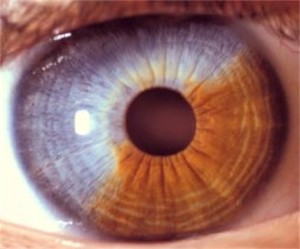
A choroidal nevus (or plural nevi) are actually moles we see on the inside of the eye, and yes, they are surprisingly common occurring in about 5% of the population.

While very common, like any mole, a small percentage of nevi unfortunately develop into a melanoma.  Because of this they must be carefully monitored.

Today we will talk about them… with lots of fun pictures


What is a Mole?


First, lets discuss what a nevi or mole actually is.  Any freckle or mole on the body is simply a benign tumor made up of melanocytes.  These melanocytes are the cells that give the skin color; darker skinned individuals have more melanocytes, littler skinned individuals less.  These normal growths develop during our lives and can be a variety of colors, sizes, and can form in groups.  If these moles become cancerous, they are given the term melanoma.


Other Types of Eye Moles


conjunctival nevusiris nevusThe human eye forms initially as an in-pouch of embryonic skin cells and since it also has melanocytes, you can also get these freckles or nevi on any part of the eye.  Here’s one on the white of the eye called a conjunctival nevus, as well as a large iris nevus.  These moles are easily seen in the mirror, what comes as a shock are the nevi inside the eye or choroidal nevus.
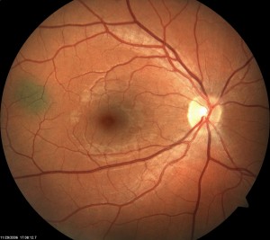
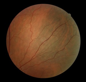

Choroidal Nevus


Choroidal nevus 1ocular melanocytosisChoroidal nevus inside the eye can come in all shapes and sizes as well.  While all nevi are by definition “normal,” they can be dark, light, small, or very, very large.  The first is a relatively small mole about 1.5 mm in size, the second photo is part of a much larger that covers about a quarter of the inside of this persons eye!

What is the Risk?


The main difference between a mole on your arm and a mole in your eye is access to monitoring it.  You could look at a freckle on your arm every day, however a mole in your eye we only get to see once a year.  The risk of a choroidal nevus turning “bad” is about 4 in 1000 and 50% of ocular melanoma develop from existing “normal” nevi.

While the risk is relatively low, choroidal melanoma is the most common primary cancer of the eye and is something we see with unfortunate frequency.  Because of this, it is important to photograph these freckles and view them through a dilated pupil every 6 months to a year.


Thanks for reading Eye to the Future and feel free to send any questions or ideas for future topics to [email protected]

Take care of those eyeballs!

Nick Wolf, OD


Giant Papillary Conjunctivitis

 Contact Lens wearers of the world, meet one of the greatest enemies of successful contact lens wear:  

Giant Papillary Conjunctivitis or GPC.

“My contact lenses wear out REALLY fast, I am constantly changing them!”

“I just can’t wear the contacts very long, they’re always moving on my eye.”

“I’m getting this weird stringy, white goop when I wear my contacts.”


What is Giant Papillary Conjunctivitis?

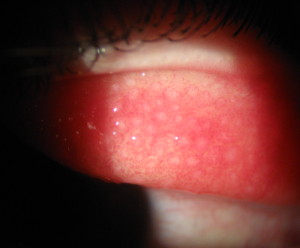
Giant Papillary ConjunctivitisGiant Papillary Conjunctivitis, or GPC as it is commonly known, is an inflammatory condition involving the conjunctiva of the human eye.

The conjunctiva is the thin slippy tissue that covers the front of the eyes as well as the inside of the eyelid.  The hallmark of this condition are small (and sometimes GIANT!) bumps called papillae that form on the underside of the upper eyelid.

This is very chronic condition and can range from mild, to moderate (such as our patient pictured left), to very, VERY severe (If you are squeamish, DO NOT CLICK!).


Holy Crap!  What Causes These Bumps?

At its core, GPC is an allergic condition and occurs almost exclusively in contact lens wearers (90 plus percent).  It can also rear its ugly head with patients (like myself) who have very severe allergies – and in the Ohio River Valley, there are lots of us!

“It is estimated that up to 35% of extended wear contact lens wearers with develop symptomatic Giant Papillary Conjunctivitis.”

With contact lens wearers every individual is different.  These bumps frequently form with certain types or brands of contact lenses, from the chemicals in contact lens cleaning solutions, or from overwear/abuse of contact lenses.

This is essentially a mechanical problem.

The brain is physiologically wired to blink the eyes every 10-12 seconds.  Every time a blink occurs the underside of the lid rubs against the contact lens.  Deposits, chemicals, and/or allergens on the contact lenses will begin to irritate the conjunctiva of the upper eyelid.  With each blink these inflamed papillae begin to form, multiply, and expand making the symptoms worse and worse.


Why do these “bumps” cause me problems?

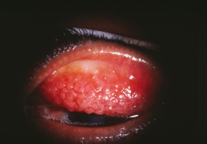
GPCThink of these papillae bumps like the grit on sandpaper.
  1. With each blink, these ‘sandpapery’ bumps rub, push and pull the lens around making it uncomfortable to wear.
  2. The bumps also poorly wet the lens making deposits form quicker.  This results in the need to change your contacts far more frequently.
  3. Finally, the areas between the bumps allow mucus to form leading to an irritating, stringy, and often blurring mucus that coats the eye.

Not to mention that as the papillae bumps multiply and grow, the rubbing on the contact lenses increases making the symptoms worse and the bumps bigger – a death spiral for contact lens wear.


What do we do about it!

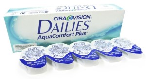
Aquacomfort PlusWell, the problem here is that when the papillae are mild, the symptoms usually get shrugged off or put up with for months or even years.

Once the symptoms are bad enough that the contact wearer come in because the contacts “aren’t working”, the Giant Papillary Conjunctivitis is usually quite ‘ground in,’ severe and chronic.

If this condition IS caught early, a simple change to Daily Disposable lenses is usually sufficient.  Daily Disposable lenses are replaced every day which means there is no time for deposits or allergens to attach to the lenses.  Additionally, there is no solutions needed for Daily Disposable lenses which removes a lot of cleaners and chemicals that contribute to the papillae bumps.

For severe problems unfortunately, there is NO simple solution and the treatment usually consists of several steps.

  1. First step is discontinuing ALL contact lens wear.  It doesn’t matter what lens we put on your eye at this point, it’s going to get manhandled by those BIG bumps!
  2. Since this is an inherent inflammatory reaction, we will start you on steroid eye drops for a period of weeks to months.  Since this is a chronic condition, it often takes a long time to reduce these bumps to a level acceptable for comfortable contact lens wear.
  3. Once the Giant Papillary Conjunctivitis is under control, we will fit you will Daily Disposable lenses to keep this from happening again.

Other treatment options for this condition included discontinuing contact lenses all together or exploring the possibility of a surgical solution to refractive error through LASIK surgery.


If you are a contact lens wearer and have noticed these symptoms in your eyes.  Please consider scheduling an evaluation with your eye care provided.  With Giant Papillary Conjunctivitis, an ounce of prevention is worth a pound of cure.


If you have any questions about this topic or any others, please contact me at [email protected]

Take care of those eyeballs!

  •  Nick Wolf, OD


Histoplasmosis – The Scourge of the Ohio River Valley

Histoplasmosis is something that most people living in the Ohio River Valley (aka the”Histo-Belt”) have probably heard about.

If not and you live the Ohio or Mississippi River Valleys, the CDC estimates there is up to a 90% chance that you have been exposed.  Given that Histoplasmosis can have severe complications to multiple organ systems, including the eyes, it’s something we think you should know more about.

Here we will look to demystify Histoplasmosis and explain:

  • What is it
  • How infection occurs
  • What symptoms it causes
  • How common is it
  • Who gets it
  • Why Histoplasmosis can cause blindness
  • How this condition is treated

What is Histoplasmosis?

Histoplasmosis actually starts as an infection caused by the fungus Histoplasma capsulatum.  This fungus is naturally found in the droppings of birds and bats and resides in the soil.  The spores of this fungus are often disturbed by construction or farming of contaminated soil and become airborne.  Exposure to Histoplasmosis occurs from inhaling the airborne spores into the lungs which becomes the primary infection site.

Acute Histoplasmosis infection in the lungs usually begins 3 to 14 days after the primary exposure and the symptoms include:

  • FeverPulminary Histoplasmosis
  • Chills
  • Chest Pain
  • Coughing
  • Muscle Aches

The severity of the initial symptoms are extremely variable.  The vast majority of Histoplasmosis infections are either symptom free or simply get written off as a mild case of the flu.  Mild to moderate cases pass without note because the body is usually able to recover within a few days to a week.  In certain cases however, like infants, the elderly, or the immunocompromised, the primary infection can be deadly and needs prompt treatment with antifungal medication.

Since the acute infection from Histoplasmosis is in the lungs, it is very common to have some evidence of a past infection, even if that infection was mild and non-diagnosed.  Many times these findings are only uncovered years later with routine imaging through X-ray or CT scans (as seen right).


Who Gets Infected with Histoplasmosis

Map of HistoplasmosisAny one, of any age, race, gender, or ethnicity is at risk for exposure and infection from the Histoplasmosis fungus.  Those of us that reside in the Histo-Belt (Ohio and Mississippi River Valleys) are at a particularly high risk.
The CDC estimates that exposure to these fungal spores in this region reaches endemic levels of as high as 90% exposure.

People at particularly high risk of infection include those in construction, aggriculture, roofing, and those who have grown up around birds or on a farm.


This is an Eye Doctor Blog, Right?

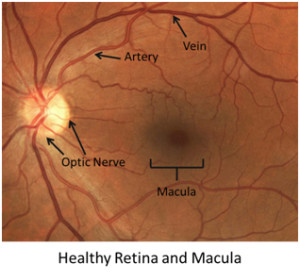
DiabeticEyeDisease-HealthyRetinaYes, yes, I didn’t forget the eyes.

While the lungs are the primary infection site for Histoplasmosis, a unknown but small percentage of patients exposed to Histoplasmosis will also develop an infection in choroid layer of their eye (vascularized layer behind the retina: healthy retina pictured at Left).

Just like the lung infection, the initial eye infection usually resolved without any treatment or knowledge that the infection occurred.  The initial infection does, however, leave a very characteristic pattern of damage.

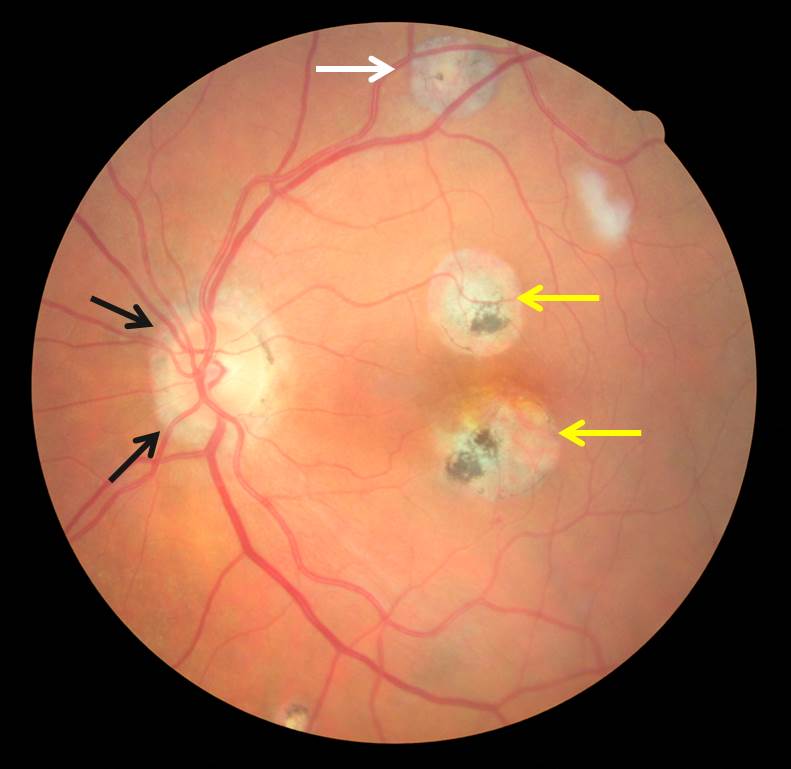
This pattern is something that eye doctors in this area see very, very often and is called the Histoplasmosis Triad.


The Ocular Histoplasmosis Triad

histoplasmosis

As the name implies, there are three distinct changes that are commonly seen in patient with a history of Histoplasmosis exposure/infection.

  1. Peripapillary atrophy or scarring around the optic nerve that leaves the back of the eye and transfer visual information to the brain (Black arrows)

  2. Punched our peripheral scars around the outside of the retina called “Histo Spots” (White arrow)

  3. Scarring of the choroid in the Macula (center part of vision where you see 20/20, Yellow arrows)

While the first two Histoplasmosis changes are benign and do not progress or threaten vision.  Scarring in the macula can be a sight threatening condition.

The risk here is NOT of infection or a reactivation of the fungus, although that is possible.  The problem here is that for an unknown reason and often times decades after the initial infection, the scarred areas at the center of vision tend to develop and grow new blood vessels.  These new blood vessels cause leaking and bleeding in this very sensitive area and require immediate treatment.  These new blood vessels develop in a similar way to Wet Macular Degeneration which was discussed in another post.

There are several forms of treatment for these blood vessel nets, I’d encourage anyone wanting to know more about the treatment to visit Bennett and Bloom Eye Center’s excellent page on this topic.


Dr. Wolf!  First you made me scared of Cats… now I’m scared of Birds…

The take home message about Histoplasmosis is that in the Ohio River Valley, exposure and sub-clinical infection is EXTREMELY common.

Most of the time, even if some ocular changes are found, there is little to no risk of progression or blindness.

What IS critical; is that if areas of scarring from Histoplasmosis are present in the center part of vision, they should be thoroughly investigated, tested, and monitored closely.

Most importantly, with Histoplasmosis so common around here, this is just yet another reason to get yearly eye exams.

Take care of those eyeballs!

–  Nick Wolf, OD

Additional Resources

National Eye Institute website on Histoplasmosis

Mayo Clinic website on Histoplasmosis

CDC website on Histoplasmosis

“Lazy Eye” and Amblyopia

Everyone has heard the term “Lazy Eye” but in reality this is not be the best term.

In actuality, it is most often not the eye that is the problem but rather the eye’s connections to the brain and the brains ability to use the eye that is really the issue.

This condition, called Amblyopia occurs in 4-5% of otherwise healthy children and is the main reason that the doctors at Precision Family Eye Care strongly recommend all children receive routine eye care to identify and treat this condition early.  Here’s why…


Amyblopia or “Lazy Eye”

As eluded to, amblyopia is most often not a disease of the eye itself, the eye is usually perfectly healthy.  However even with the correct glasses prescription in place, the eye does not see things normally and is termed “lazy”.

How can that be??  

Well, when a child is born there is no vision in the womb, therefore there is very little vision at birth.  Like walking, talking, and crawling, vision is learned.  It is only after an infant starts examining the world around it that firm hard wiring begins.

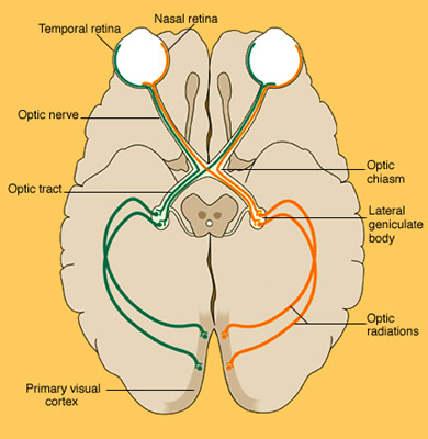
visual-pathwayThe eyes are obviously in the front, but they are a camera, nothing more.  What actually “sees” is the occipital lobe at the back of the brain.  The pictures from the eyes are sent via the optic nerve to the Lateral Geniculate Body (LGN) in brain for processing.  After that, the processed signal is sent by optic radiations called the Meyer’s Loops to the Primary Visual Cortex of the occipital lobe which actually interprets the picture (diagram right).
Because this complex pathway requires active stimulus or pictures from the eyes to form, this process can be disrupted if there is not a normal picture coming from each eye. This process of abnormal visual development can occur in one or both eyes and for a number of reasons.  The most common reasons for Amblyopia are high or asymmetric eye prescriptions and strabismus (eye turns).

If found early, treatment and training is available to help build this nerve network; if not, the vision reduction is permanent.


Types of Amblyopia

Refractive Amblyopia

Refractive amblyopia occurs when there is a large or very unequal refractive error (glasses prescription) present at birth. Even though the eyes are healthy, if one or both eyes are unable to send a clear image to the brain, the brain will not connect to the eyes properly.  One of the most common scenarios is when one eye is normal (little or no glasses prescription required) and the other eye is born very farsighted. In this scenario, a child will see clearly with both eyes open and rarely complain of problems because their brain is only paying attention to “the good eye.”

Pediatric Strabmismus (Eye Turn) Amblyopia

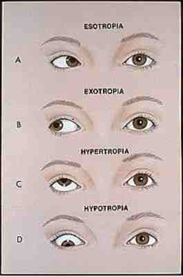
strabismusStrabismus is a condition that occurs when the eyes aren’t properly aligned with one another from birth.  This results in one eye turning in, out, up or down with respect to the other eye. When the eyes are not working together properly, the brain is getting two different pictures from the eyes.  This leads to double vision which the brain does not like.  As a result, the brain will then decide to pick only one eye to develop properly and the input from the other turned eye will be ignored.

Without the input from that eye to develop the needed connections to the brain explained above, the turned eye will have permanently reduced vision if not properly diagnosed and treated.  Eye turns can be quite obvious, however, amblyopia can occur from even mild eye misalignment that often goes unnoticed by parents and teachers.

Both of these types of Amblyopia often result in only one eye being affected and both often go unnoticed without a proper eye exam due to one eye developing properly and the child not understanding that only seeing out of one eye isn’t “normal.”


Treatment of Amblyopia

PediatricEyeCareRegardless of the cause, is important to identify and treat amblyopia as early in life as possible.

If the development of one or both eyes is stunted, it is not only possible, but probable, that vision will be improved with treatment.  The treatment of amblyopia is directed to improve vision by strengthening the brain’s connections to the weaker amblyopic eye. This can be done with a combination of full time glasses, wearing an eye patch, or sometimes eye drops.  As the brain pays more attention to the amblyopic eye, the essential connections between the eye and the brain can be improved, resulting in better vision for that eye.

The key to treatment is timing!!

Children posses amazing neuroplasticity or the ability to rework connections within the brain.  Just like everything else though, humans get stuck in our ways over time, and our brains are no different.  The visual pathway continues developing from birth through age 7 – 8.  If treatment is given before this time, the prognosis of vision improvement is very good.  After age 8 however, amblyopia may be resistant to treatment as the visual pathway has already completely developed.


The take home message here is that “Lazy Eye “or Amblopia is more of a brain problem than an eye problem.  However, this is very treatable, but requires a proper early diagnosis.

Therefore, all children should receive an eye exam by age 3 and then every year as they go through school ages to ensure them the best chance at a lifetime of healthy eyes and excellent vision.


As always, feel free to contact us with any questions or ideas for future topics.

Nick Wolf, OD

For more information on Amblyopia, vision development in children, and binocular vision problems please visit our website pages or visit the National Eye Institute’s excellent page about this condition.