Contact Lenses of the Future


Contact Lenses for more than Vision


As optometrists, we fit contact lenses daily.  While new contact lens materials and designs have greatly increased the comfort and vision of our contact lens patients, we couldn’t help but wonder if contact lenses had additional diagnostic potential…

With that backdrop, some recent announcements and advancements in contact lens technology has got us very, very excited.  Several different companies are now looking at ways of incorporating recent technological breakthroughs into contact lenses for the purposes of disease monitoring.

Today on Eye to the Future, we will delve into two contact lenses of the future.


Google/Alcon’s Blood Sugar Monitoring Lens

Google Contact LensThe first, and one you might have heard about, is a recent paring between Google and Alcon (eye care branch of the drug company Novartis).

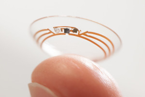
While I personally cannot wait for a self driving car, the geniuses over a Google have equally excited our profession with their recent announcement to use their expertise to develop a contact lens that monitors blood sugar for diabetic patients.

This contact lens (pictured at left) would contain micro sensors to measure the blood sugar levels in the tears as well as a radio antenna thinner than a human hair.  The data is then transmitted from the antenna to a smart phone app to allow the continuous monitoring of blood sugar.

With the epidemic of diabetes in this country, this non-invasive contact lens of the future could revolutionize the way patients monitor their blood glucose levels.  In addition to the easy of measurements and lack of finger sticks, this information would be enormously helpful for primary care doctors and endocrinologists in developing specific treatments plans depending on how blood sugar levels change throughout the day.


The Triggerfish by Sensimed

Sensimed TriggerfishEveryone who’s been to the eye doctor knows the dreaded “Puff of Air” but what that test is actually measuring is the pressure inside your eye.

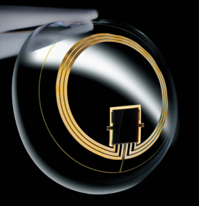
The Swiss company Sensimed has developed a wearable tech heavy contact lens called the Triggerfish.  This contact lens of the future (right) was designed for the sole purpose of continuously measuring eye pressure over a 24 hour period.

Eye pressure, or intraocular pressure (IOP), is an important factor in the diagnosis of Glaucoma and even more important in determining the effectiveness of a Glaucoma treatment.  The problem with eye pressure is that it fluctuates quite a bit throughout the day, as much as 50% or more in glaucoma patients.

IOP tends to be lowest in the afternoon or early evening when many people get eye exams.  This can give us a false sense of security that the eye pressure is “normal”.  Even worse, the IOP also tends to be highest when we can’t record it, usually the very early morning hours when most of us are just waking up or enjoying our morning cup of coffee.

Having a graph of the high and low points with this non-invasive testing would allow eye doctors the ability to choose specific treatments that are most effective when the eye pressure tends to be highest.

Triggerfish worn
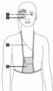
The Triggerfish is worn in one eye (1).  A wireless antenna (2) is positioned around the eye to collect the data from the contact lens.  Then a set of wires (3) sends data from the antenna to a recording device worn by the patient (4).

The patient is fit with the contact lens and antenna in office and simply wears this device as well as a monitoring pack for 24 hours before returning to the doctor where the data is downloaded and analyzed.  This is very similar to heart monitors often used by primary care doctors and cardiologists to record a patients cardiac measurements outside the office setting.

This lens is currently going through studies in the US to determine its safety and effectiveness, but is already in full use overseas.

For additional information on the Triggerfish diagnostic contact lens, please visit the Sensimed website.

We hope you have enjoyed this little overview of the some contact lenses of the future.  If you have any questions, as always, please contact us and ask.

–  Nick Wolf, OD

Patient Question – “What are Floaters?”


What are Floaters?

floaters

The technical term for these visual phenomena are called Muscae Volitantes which is Latin for “Flying Flies.”  No one refers to them that way though, they are simply called floaters.  Commonly floaters are noticed as specks, strands, or ‘ameba-like’ see through forms that drift through the vision from time to time.  They will often move as the eye moves and appear to change shape as they float about.

eye dilationThe first important thing to know is that floaters come from, and form in, the jelly of the eye called the Vitreous (right).  Floaters can form at any age and can range from mild to severe.

The jelly inside the eye is quite thick and gel-like as a child but over our lifetime it breaks down and liquefies.  As the jelly liquefies, it gets sticky and clumps begin forming.

These floaters are suspended within the jelly of the eye and with eye movement they become “shook up” and drift about similar to snow floating inside a snow globe.  As these clumps pass through the center part of vision you will see the shadows of them.

Floaters tend to more visible against a flat background, like a bright blue sky or a freshly painted wall.  Additionally, floaters are more pronounced in bright environments because bright lights make the pupil smaller which casts a more visible shadow from the floater onto the retina.

Below is a fun and quite educational video explaining floaters (as well as another commonly described and really cool “eye quirk” called the Blue Field Entoptic Phenomenon… that’s so cool it deserves a blog post of it’s own).


Should I get checked if I see Floaters?

While hugely annoying, floaters in general are a normal and mostly benign condition.  It is important to note however, that any new or changing presentation of floaters should be examined for a potential eye problem.

As anyone who’s gotten a corneal abrasion can attest to, the surface of the eye is highly enervated and sensitive.  By contrast, there are no nociceptors (or cells that sense pain) inside your eye.  Therefore, if you develop a problem on the inside of your eye like a tear in the retina, a bleed, or an infection, you will feel no pain; your only symptoms will be an increase in the floaters you see.

Additionally, floaters in conjunction with bright flashes or arcs of light in the side vision, is cause for concern and should be checked.  Flashes of light come from traction of the jelly on the retina and could lead to a problem.


One Floater to ALWAYS get checked

posterior-vitreous-detachmentOne specific type of floater that should always be investigated is something called a Posterior Vitreous Detachment (or PVD for short).  PVDs occur as the liquefying jelly in our eye begins to collapse in on itself and peels away from the lining inside the eye, called the retina.

This process is a near guarantee with age, occurring in about 50% of humans at age 55, up to 86% by 90.  When the vitreous, or eye jelly, pops off its attachment to nerve in the back of the eye, it leaves a very large, very annoying circular floater right in the center of vision.  This floater is referred to as a Weiss Ring (pictured left).

The problem here is not the floater itself.  Instead, as jelly peels away from the retina, it will often tug on the retina and has to potential to cause retinal tears in up to 10% of patients with an acute PVD.


Is there a treatment for Floaters?

The vast majority of the time floaters are a normal, albeit irritating, condition.  Since these little buggers are actual inhabitants inside the eye, there is no simple treatment to remove them.

One option is something called a vitrectomy, where a retinal surgeon removes the jelly of the eye and replaces that jelly with saline.  This is a significant operating room type surgery with lots of potential complications and therefore is rarely performed for floaters alone.

A newer treatment option that has surfaced in the last decade is to use a high powered laser to essentially “blow up” the floaters.  While a novel approach, putting this much laser energy into an eye has the potential for very serious complications making this a very questionable approach.

What we do have going for us is that over time the brain will learn to “see” floaters less and less and gravity will also take it’s toll and floaters will sink out of vision.

Bottom line, the best treatment for floaters: try to ignore them.


We hope this discussion has answered some of the questions you had about Floaters.  If you think of any other questions, feel free to let me know!

Take care of those eyeballs!

–  Nick Wolf, OD

For additional reading material on Floaters, please visit the Mayo Clinic’s excellent page.

What’s New – Kamra Corneal Inlay


Kamra corneal inlay by AcuFocus, Inc.


Some things in life are annoying and unavoidable, reading glasses certainly fall into that category…

Up to this point, there really has not been a surefire, surgical way to fix the near problems associated with presbyopia or the “Curse of the 40s.”

conductive keratoplasty
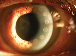
Conductive Keratoplasty (pictured left) was a procedure that was done in the past by making thermal burns in a circular pattern on the cornea… this was pretty much a resounding failure and this procedure is rarely performed anymore.

Another option was and still is LASIK.  Near vision with LASIK is achieved by surgically inducing monovision, where one eye is corrected for distance and the other for up close.  Think of it like the 21st century version of the monocle and is commonly done with contact lenses.  This is an effective correction option but tends to become less useful for near vision over time.

The best surgical option currently available is doing cataract surgery and putting in a lens that can actually focus for distance AND up close.  Cataract surgery without a actual cataract is considered a cosmetic, out-of-pocket cost by insurance companies.  With the add on cost of a couple of these high tech lenses, the price tag quickly becomes cost prohibitive to many individuals.


Introducing Corneal Inlays


karma corneal inlayCorneal inlays are small pieces of plastic that are installed inside a pocket in the cornea.  The pocket is created with the same laser that is also used to make the “flap” during LASIK surgery.  The inlay is slid into the pocket, positioned correctly, and then the pocket seals itself.

There are several cornea inlays used for presbyopic correction overseas and in Canada and a couple are slugging through the FDA approval process in the US.

The one most likely to be available to bifocal wearing Americans in the near future is the Kamra inlay by AcuFocus, INC.

The Kamra corneal inlay (shown above) is about 1/5 of the size of a contact lens and is implanted into only one eye.  This plastic inlay has one central hole and over 8,000 tiny holes in the plastic to allow nutrients to flow through.

This device works by using the pinhole effect.  Not to get too far into the weeds here, but if you every played with a pinhole camera as a child, or are familiar with photography, you know that making a smaller opening (or f-stop) on a camera increases the depth of focus.  The Kamra corneal inlay only allows light to pass through a very small 1.6mm central hole and not through the normal pupil size which ranges from 3-7 mm.  This greatly increases the available ranges that the human eye can focus on, particular with reading and computer tasks.

Since this device is positioned over the pupil, it is invisible to the naked eye.  The video below shows several different patients with the kamra corneal inlay.  It also does not effect the distance vision but allows for some additional vision for near tasks without reading glasses or bifocals.  Unlike the other surgical options discussed above, this inlay can also be removed if treatment is not effective.

https://www.youtube.com/watch?v=GKECqj0JiDQ


This product is still in the investigatory stage and will likely not be available to patients (outside of research centers) until 2016 or later.  For more information please visit the AcuFocus, Inc website about the Kamra corneal inlay.

We hope you have enjoyed this walk-through of a new technology you may be offered in the future and we will continue to keep you informed of any new or interesting advancements as we keep an Eye to the Future.

Take care of those eyeballs!

– Nick Wolf, OD

That’s Real?!? – Charles Bonnet Syndrome

“I see tiny men with hats” aka Charles Bonnet Syndrome

Severe vision loss and blindness in the United States is an unfortunate reality.  Conditions like macular degeneration and diabetic eye disease are often the culprits.  When blindness does occur, a significant number of patients will inexplicably begin seeing brilliant, colorful, vivid, and reoccurring visual hallucinations all while being otherwise perfectly sane.  This bizarre and often unsettling condition is referred to as Charles Bonnet (“bō-NAY”) Syndrome.

Charles Bonnet SyndromeWho Was Charles Bonnet?

Charles Bonnet was a Swiss philosopher who, in 1760, first described this phenomenon that would later bear his name.  Bonnet’s grandfather was nearly blind in both eyes from cataracts (long before surgical repair was possible).  Even with the visual impairment, his grandfather often described “seeing” complex scenes, people, animals, and even buildings and landscapes.  Although Bonnet was quick to understand the link between blindness and visual hallucinations, Charles Bonnet Syndrome was not officially recognized as a defined psychological condition until 1983.

How is “vision” still possible with blindness?

The visual hallucinations seen with Charles Bonnet Syndrome are obviously not real but simply phantom visions.  The brain desperately wants to continue using the eyes for visual stimulus.  However when eye disease robs the brain of that ability… the brain essentially… makes stuff up!  The process is quite similar to phantom limb syndrome where sensations, pain, or tingling are often noted in a previously amputated limb that is no longer there.

What do sufferers commonly see?

Charles Bonnet Hallucinations
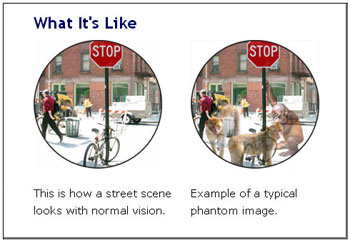
Visual hallucinations from Charles Bonnet Syndrome are quite varied but most often involve patterns, animals, people, or scenes and commonly fit into the individual’s actual surroundings.  One characteristic of Charles Bonnet visions is that they are commonly “Lilliputian” hallucinations meaning everything and everyone look much smaller than they would normally appear.  This trait of Charles Bonnet hallucinations results in individuals commonly seeing small pixie-like figures of varying colors and appearances.

These hallucinations usually occur during down time and not while the patient is active.  Additionally, these hallucinations are visual only and do not involve interaction, smell, sound, taste, nor touch and usually are actually quite soothing and non-threatening when they occur.

This.  Is.  Normal.

Yes, visual hallucinations from Charles Bonnet Syndrome are in fact normal and occurs in otherwise completely mentally healthy individuals.  Suffers realize that these visions and images they are seeing are not real.  Actual statistics for Charles Bonnet Syndrome are quite difficult to fully assess.  This is not surprising as individuals who experience these hallucinations are hesitant to discuss them with friends, family, or doctors for fear of stigma or being labeled as mental insane.  It is estimated by most researchers to be extremely common affecting anywhere from 14-40% of individuals with recent blindness.

There is also no treatment for Charles Bonnet Syndrome.  These visions occur off and on for the first 12-18 months after blindness but usually become less and less frequent over time.  The most important treatment is to educated patients that although this condition can be unsettling, it is a normal neurological response to recent blindness and not a sign of mental illness.

Feel free to follow up with any questions or comments and thank you, as always, for tuning into Eye to the Future!

  • Nick Wolf, OD

For more information on Charles Bonnet Syndrome and/or talking about this condition with blind family members please visit the Royal National Institutes of Blind Peoples’ excellent page about this condition.

Glaucoma Awareness Month

glaucoma

Glaucoma Awareness Month – Q & A

Glaucoma is a condition that most everyone has heard of, but quite a bit of confusion still exists.

Since January is Glaucoma Awareness Month, we will be doing a Q&A style post giving some simple facts and debunking some myths about Glaucoma.

If there’s a question I missed feel free to ask.  For a far more in-depth discussion on Glaucoma, please visit our glaucoma webpage or the National Eye Institute’s page about Glaucoma.


What is Glaucoma?

Glaucoma is a disease where the internal pressure of the eye causes irreversible damage to the optic nerve that leaves the back of the eye and carries the visual signal to the brain.


How common is Glaucoma?

It is estimated that Glaucoma afflicts about 2.5-3 million Americans… the scary part is; only half of them know they have it!


Does family history play a role?

Absolutely!  In the Barbados Eye Study, having Glaucoma in your nuclear family (parents and siblings) increased your risk of developing Glaucoma to as high as 20%.  Furthermore, African or Hispanic ancestory also increases the risk 4 fold.


But I don’t feel eye pressure problems?

Unlike high blood pressure which makes you feel funny when elevated, high eye pressure is rarely ever felt unless it gets very, VERY high.  This means you have no idea that your own eye pressure is higher or lower than normal without testing.


My eye pressures are always normal at the eye doctor, so I don’t have Glaucoma?

Not so fast.  The Baltimore Eye Study sought to answer this question, looking at hundreds of new patients with Glaucoma and looking for trends.  In that study, 55% of newly diagnosed Glaucoma patients had normal eye pressure at their first visit and 16% had normal eye pressures throughout the whole study!  So no, just because your eye pressure is at a “normal” level doesn’t mean you are safe from this eye disease.


I have been told I have high eye pressures, which must mean I DO have Glaucoma?

No, as well.   Many people fall into the category called “ocular hypertensive,” meaning the eye pressure is slightly increased compared to what is considered normal but no active damage is occurring to the optic nerve.  A study called the Ocular Hypertensive Treatment Study (OHTS) looked at the risk involved with elevated eye pressure.  While high eye pressure is surely a risk factor for Glaucoma, that study found that only 10% of ocular hypertensive patients developed glaucoma damage after 7 years of no treatment.

Therefore, if your pressures are elevated it is important to monitor closely and treat if necessary, but not all individuals with high eye pressures need treatment.


Well if eye pressure doesn’t tell you if I have Glaucoma, what does?

Not a simple question.  Diagnosis of Glaucoma is like putting a puzzle together, and your eye pressure is only one piece.  In order to fully rule out Glaucoma, some additional information is needed to evaluation your optic nerve health, eye drainage, and peripheral vision among other things.  This information is collected with simple, painless in office testing.


How does Glaucoma affect vision?

glaucoma vision loss

Glaucoma vision loss is devious.  Unlike other eye diseases that cause loss of central vision, Glaucoma causes side vision loss.  Peripheral vision loss from Glaucoma is impossible to perceive early in the disease.  By the time enough side vision has eroded to become obvious to the patient, you have lost up to 90% of your vision and the disease has reached irreversible end stage with limited treatment options.

That coupled with the lack of actual symptoms from eye pressure has led to Glaucoma being called the “Sneaky Thief of Sight.”


Can it cause Blindness?

Unfortunately yes.  Glaucoma is the third leading cause of new blindness in the United States and the second leading cause of blindness worldwide.


I have Glaucoma, what is MY risk of blindness?

A study was once performed where glaucoma was diagnosed and simply followed, not treated.  In that study, 29% of those individuals reached blind, end stage disease in 10 years.  However, with early diagnosis, proper treatment, and comprehensive monitoring, the risk goes down to only 6% over 15 years.


How is Glaucoma Treated?

While eye pressure alone is not a sure sign that Glaucoma is present or absent, study after study has shown that lowering eye pressure prevents further vision loss.  Therefore, the main treatment for Glaucoma is to lower the eye pressure through the use of prescription eye drops or some surgeries in advanced cases.


Since Glaucoma is such a silent disease.  The key to keeping this condition in check is routine, preventative care.  If you have any risk factors for Glaucoma outline on our website, it is highly advised to receive at least a yearly Comprehensive Ocular Health Exam.

As always, please email with additional questions and take care of those eyeballs!

–    Nick Wolf, OD

Bifocal Contact Lenses

Contact Lens FittingThe technology of contact lenses has come a long, long way in the past 10 years, solving most of the problems that people associate with these medical devices.

Contacts lenses irritate my eyes”… Done!… more natural and wettable materials to improve comfort!

I can’t wear contacts because of my astigmatism”… yes you can with new toric technology!

But I’m over 40 and need to wear reading glasses with my contact lenses”… well, let me tell you about Bifocal Contact Lenses…

Bifocals contact lenses aren’t necessarily new to the contact lens market.  The first bifocal contact lenses came out about 20 years ago; however, it was so darn bad that it turned off a lot of patients (and doctors) to the potential of these designs.  As manufacturing techniques and technology have improved, this class of contacts has reaped the rewards.  Bifocal contact lenses these days are stunning, giving patients clear distance and reading vision with an 85-90% success rate.

How do Bifocal Contact Lenses Work

The key to bifocal contact lenses is in the design.  These lenses are bifocals in the name only and work nothing like a pair of bifocal glasses.  Bifocal glasses have different zones within each lens with very different prescriptions for clear vision from distance to near.  You simply need to direct your eyes through that section of the lens for clear vision at that whatever distance you want to see.  Since a contact lens is so small and always sits at the same place on the cornea, it is not feasibility for the lens to move around enough to look through different areas.

Bifocal Contact Lenses
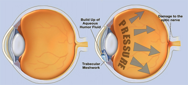
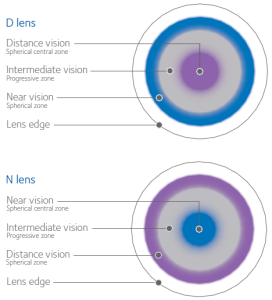
Instead, if you look at these lenses under a microscope they have concentric rings just like the rings on a dart board.  Each ring of a bifocal contact lens is designed to focus for a different place: distance, intermediate or computer, and reading (see right).  All the vision from all distances is focused by the contact onto the retina in the back of the eye and the retina sends that information to the brain.

What the brain receives is a garbled, distorted mess, but the supercomputer sitting between our ears is able to filter out the distances that it’s not interested in.  This results in you being able to read a menu one moment and then check the score of a ball game on a television the next.  This process sounds quite convoluted but it actually works surprisingly well!

Additionally, there are multiple parameters we can change and adjust to fine tune the vision through these contacts based on how they work for the patient in real life situations.

Keys to Success with Bifocal Contact Lenses

There is no panacea, or cure all, for the “Curse of the 40’s” but Bifocal Contact Lenses are a large step in that direction.  The keys to success with these lenses are realistic expectations and patience.  Since the brain has never used lenses like this before, it takes 2-4 weeks of daily wear for the brain to understand how these lenses ‘work.’  During that time it is common to notice some distortion and blurring but most of that goes away over time.

If you are new to Bifocal Contact Lenses there has never been a better time to try out this technology.

Schedule an appointment with you eye care specialist today!

–  Nick Wolf, OD

That’s Real?!? – Cat Scratch Fever

 This will be a recurring series on Eye to the Future, where we will be looking into some of the more peculiar and odd eye conditions out there.  Hopefully you will find these topics interesting, and if nothing else, maybe you can astonish your friends and family during Jeopardy!

ted nugentYes!!!  Amazingly enough…

In addition to being a thinly veiled sexual euphemism made famous by rocker Ted Nugent, Cat Scratch Fever (or more accurately Cat Scratch Disease), is actually a relatively common condition that can cause some substantial eye related problems.

Cat Scratch Disease is a constellation of various systemic (and eye) symptoms that results from, logically, a cat’s scratch or bite.  The underlying cause of Cat Scratch Fever is a bacterium called Bartonella (below, right) that was confirmed by Robert Debré in 1950 to be naturally occurring in cats and potentially infectious to humans.  Bartonella is more commonly found in kittens/young cats and is most likely to occur in outdoor cats as fleas are the main method that Bartonella is spread within cat populations.  Cats pick up this bug through fur cleaning of flea debris and foster this bacterium in their saliva which results in the spread to us through bites and scratches.

bartonellaWhen Bartonella is transmitted to humans it has a latent period of 1-3 weeks from the initial bite, but it is not uncommon for symptoms to only pop up 1-2 months later when the memory of the cat scratch has long faded.  Cat Scratch Fever commonly presents with tender, swollen lymph nodes near the site of the scratch/bite and the afflicted also suffer from severe flu like symptoms with malaise, aches, fever, headache, and joint/muscle pain.  In very severe cases, Bartonella can also can meningitis (infection/inflammation in the brain) or endocarditis (infection/inflammation in the heart).

Patneuroretinitis
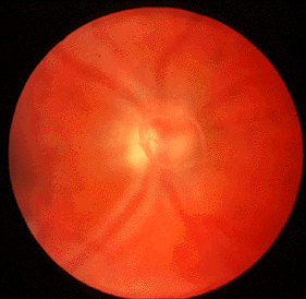
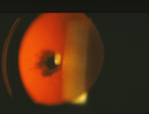
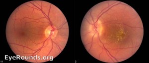
ients with undiagnosed Cat Scratch Fever will often make a trip to their local eye doctor during this time because Cat Scratch Fever can cause a severe conjunctivitis (pink eye) and even very blurry vision from optic neuritis or neuroretinitis (inflammation of the optic nerve and retina).  The optic nerve carries the signal from to the eye to the brain and inflammation of this nerve results in decreased vision (left photo is normal, right has an inflammed optic nerve and retina).  The blurry vision from Cat Scratch Fever neuroretinitis is usually temporary.

Now, take a breath…. I know you are likely glancing at your friendly, family feline with a mixture of repulsion and horror…  Fear not!  Cat Scratch Fever from Bartonella is usually very benign and all symptoms typically abate within a couple weeks without any treatment.  In fact, Bartonella infection is estimated to be quite under-diagnosed simply because severe complications are rare and symptoms are similar to the flu, and like the flu, improve on their own.  If severe complications from Cat Scratch Disease do present, it is possible to shorten the length of symptoms through oral antibiotics, but the medical literature is conflicted on which medicines work best and how much treatment actually improves the condition.

I hope you’ve enjoyed this quick talk and the next time you hear Nugent’s risqué anthem playing on the radio… let it be a reminder to think twice before petting stray cats!!

–  Nick Wolf, OD

What is Cataract Surgery?

Last time around on Eye to the Future, we discussed in detail What is a Cataract?.  This time around we will be addressing what we do about it!  Mainly, cataract surgery.

As discussed before, it is common for clouding of the human lens to be present on most individuals over the age of 50; this condition is called a cataract.  However, it typically takes a long time (as in over a decade or more) to reach the point where the human lens has clouded enough to require surgical intervention.  It is possible to tweak glasses prescriptions, improve lighting, and use anti-reflective coatings on the lenses to cut down on the symptoms associated with cataracts.  However, the best method for addressing this condition head-on is through cataract surgery.

cataract before and after
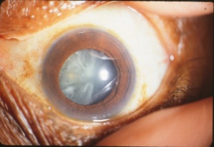
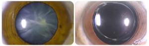
Cataract surgery permanently removes the clouded cataract and replaces our human lens with a crystal clear, man-made lens.  Above is an advanced cataract on the left and the same eye after  the cataract is removed and the implant positioned.  It’s not hard to imagine the improvement this will have on vision.  And the way in which this surgery has evolved as well as some newer implant options available is just…. awesome!

It should also be clearly stated that there have been no peer reviewed, credible studies to suggest, support, or confirm that any other treatment exists (supplements, vitamins, medications, etc) to improve the symptoms or severity of cataracts – other than cataract surgery.

Cataract Surgery Procedure

cataract surgery
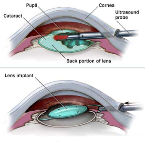
Cataract surgery is an outpatient procedure performed under general anesthesia which means you are awake but sedated.  Cataract surgery is done one eye at a time (separated by one or more weeks of healing).  During this procedure a cataract surgeon will make two small incisions in the cornea  to allow instruments to be inserted into the eye.  The crystalline lens sits in a thin membranous bag and to get to the lens, the front surface of the membranous bag is removed.  In the past, the lens was removed whole, requiring a very large incision and a long recovery time.  Nowadays, the surgeon uses an ultrasound probe to break up and remove the lens inside the eye.  Now that the clouded lens is removed, a man made lens is put back in it in place allowing clear vision once again. The incisions are self-healing and no stitches are required. The entire surgery takes about 10 minutes.

Below is a great video of a routine cataract surgery as well as easy to understand narration of the steps.

Disclaimer for the squeamish:  There is little to no blood, but this is a video of a surgery.

Cataract Surgery Risks, Recovery, and Rewards

All surgeries should be taken seriously, however, cataract surgery is the most frequently performed outpatient procedure in the United States with a very low risk for permanent vision complications (only about 1-2%).  The standard post-operative for monitoring is usually one month. During this post-operative period, eye drops are used frequently to prevent infection and inflammatory complications and follow up visits are required in order to check on the healing process.

Recovery is usually rapid with substantial vision improvement within a few days of the operation.  A large study recently found that 95% of adults were satisfied with their vision and quality of life after cataract surgery.  Furthermore, having cataract surgery also decreased the risk of car accidents by 2.5 times and serious falls and fractures by 34%.

Cataract Surgery Implants

RestorWhen the cataract is removed from the eye, there is an opportunity for the surgeon to put any powered lens back in its place.  Measurements are taken before the surgery to measure what powered lens is needed to correct for any underlying prescription like nearsighted, farsighted, or astigmatism.  A lens is then chosen to correct for your individualized glasses prescription which results in most individuals being far less reliance on distance glasses after cataract surgery.

Even more exciting is that in the past decade, more advanced lens implants have come to market like the Restor Lens at right.  In addition to making distance vision clear, these lenses can also focus for up close reading tasks as well!  While not a perfect system, this type of lens up-grade can actually free patients from all glasses for most activities!!

All surgeries can be stressful, scary, and uncertain times, but at Precision Family Eye Care we see cataract surgery as an opportunity and only work with the very best surgeons in our area to give our patients a comfortable experience and a rewarding outcome.

As always, if you have any additional questions.  Please feel free to contact us.

–  Nick Wolf, OD

What is a Cataract?

cataract diagramWhat is a cataract?  Are there different types?  Why does it form?  What are the symptoms?  What can you do about it?  All good questions that eye doctors get asked on a near daily basis.  Today on Eye to the Future we will seek to answer these and more as we demystify Cataracts.

A cataract is the most common ocular disease that results in decreased vision and usually occurs from the normal aging of our eyes.  Inside our eyes, we have a clear, round lens (left) that is used to focus light onto the retina.  When this lens is clear the light passes easily through.

Over time, however, this lens becomes cloudy and yellowed resulting in a cataract.  This change results in two problems: First, the amount of light that is able to pass through the cloudy lens is decreased so the eye continues to need more and more light to function.  Second, the light that is able to pass through the cloudy lens bounces off the cloudy areas and is scattered, resulting in increased glare and starbursts around lights.

Common symptoms that are observed when a person has a cataract include: generalized blurry vision, difficulty distinguishing colors, glare (especially at night), difficultly seeing to read books, and having to use more and brighter light to read up close.

Common Types of Cataracts

nuclear cataractNuclear Sclerotic cataracts are by far the most common and occur from a yellowing of the lens (seen at right).  This change is normal and occurs naturally with age, although certain things like smoking can increase the speed of development.  Although some yellowing of the human lens is often seen as early as age 50, it usually takes a decade or two for this type of cataract to cause visual problems.

cortical cataract

Cortical cataracts look very similar to spokes on a wagon wheel.  These cloudy spokes begin at the outside parts of the lens and grow toward the center causing visual problems.  This cataract is also common with age and is highly associated with systemic Diabetes.  Cortical cataract also develops more quickly than Nuclear Sclerotic cataracts.


posterior subcapsular cataract

Posterior Subcapsular cataracts are less common.  This can be a normal aging change, however this type is most commonly associated with long term oral steroid use or a history of past eye surgery.  This type of cataract is a cloudy area on the back of the lens and tends to develop the fastest and cause the most vision problems of all three types.


Treatment of Cataracts

Regardless of the type, when cataracts are first forming it is common to simply monitor them every so often and record their progress.  However, over our life time, our cataracts will often get to the point that light is no longer able to get through that clouded lens, resulting in permanently decrease vision.  Furthermore, there is little that new glasses will help with, because the problem is inside the eye.  At this point, the only option available to improve vision is cataract surgery.

I hope this has dispelled some myths about cataracts, next week we will be going into detail on cataract surgery.  What is involved?  How is it done?  What are the risks?  We’ll even include a video of an actual surgery!

As always, please do not hesitate to contact me with additional questions and we’ll see you next time!

–  Nick Wolf, OD

American Diabetes Month

In this connected day and age, everyone reading this knows about the epidemic of Diabetes worldwide.  November is American Diabetes Month and I wanted to bring additional awareness because the statistics from the CDC in the US alone are stunning…

9.3% of the US population has diabetes and over 1/4 of those are undiagnosed cases

28% of the US population is pre-diabetic with a strong likelihood of progression

The projected statistics suggest that this epidemic of diagnosed Diabetes may reach 25% by 2050

The total cost of Diabetes to our economy and health care system in 2012 was $245 billion (yes that is in billions, with a B!)

In addition to health risks like kidney disease, heart disease, stroke, and amputations; Diabetes is also the number one cause of new blindness in the United States.

Diabetic Retinopathy Statistics
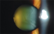
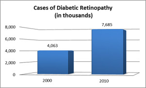
Even more concerning to eye care providers is that the incidence of ocular changes in Diabetic patients has increased by 90% in the past 1o years.  This percentage outpaces even that of new diabetic diagnoses and suggests that the blindness rates from Diabetic complications will only continue to rise…

While these are grim statistics, there is a substantial silver lining.  While the rates of blindness and eye complications have risen, eye care has also evolved greatly with better ocular testing equipment and vastly improved treatments for diabetic complications of the eye.  The key to preserving vision is earlier diagnosis of eye problems, close monitoring, and better patient understanding of what eye problems can occur and lead to blindness.

If you or a loved one has Diabetes it is absolutely imperative that you receive yearly dilated eye exams with an eye care provider.  Schedule your exam today!!

For a far more thorough discussion on the eye complications that can arise from Diabetes please visit our webpage on Diabetic Eye Disease.

Please take care of those eyeballs!

– Dr. Wolf