Dr. Wolf’s – Contact Lens Care Tips

Contact Lens Fitting
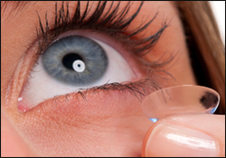
Every contact lens wearer remembers their first experience with contact lenses- the excitement, the freedom from glasses.  However, what is often forgotten is the tips, training, and recommendations given by the eye care provider.  Today we will be doing a quick refresher on proper contact lens care.

Why is this even necessary?

Simply put, getting into the habit of proper contact lens care goes a very, very long way to ensuring you have a comfortable and healthy experience while wearing these medical devices.

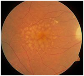
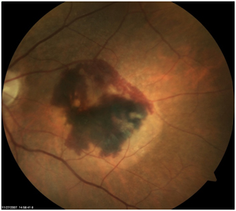
I could put up picture after picture of the horrible things I’ve personally seen that were caused by contact lens misuse and abuse.  However, let’s just say that complications from contact lenses are a leading cause of preventable blindness in US.  If you’re inclined to horrible graphic pictures, type “contact lens corneal ulcer” into google images…

We don’t like seeing these complications so here are the recommendations for contact lens care that we try to instill at Precision Family Eye Care:

  • Always wash and dry your hands before handling the contacts and avoid moisturizing soaps that will leave a film.
  • Contacts come first – put in the contacts before applying makeup or creams to prevent cosmetics from adhering to the lens.
  • Use only the multipurpose cleaning solutions recommended by your doctor.  Eye drops, saline, or tap water will not disinfect your contacts and will cause a problem.
  • Please rub to clean the contacts.  Rinsing and soaking is not sufficient.  A simple cleaning will go a long way to keeping the lenses fresh and clean.
  • Never, ever reuse or top off contact lenses solution!!  This cannot be overstated.
  • contact lens case
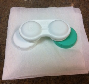
Each time you remove you contact lenses from the case you should dump the solution, rinse the case, and allow it to fully air dry before the next night’s use.  We recommended this position for the case throughout the day (picture right).  A closed case will not air dry and grow bacteria and a face up case will collect hairspray.
  • Even with perfect care, a case will begin to grow bacteria over time.  Cases should be boiled or replaced at least every 3 months.
  • Please do not wear your contacts while swimming.  Chlorine is very rough on contacts and there are plenty of critters in both hot tubs and fresh water that would love to start growing on your eyes.
  • Sleeping over night in contacts increases your chances of getting an eye infection by 15 times!  If you absolutely must, please let you eye care provider know so they can prescribe lenses made for overnight wear.
  • Contact lenses carry a FDA approved wearing time for a reason.  If you use eye irritation as the judge of when you change your lenses, you are already doing microscopic damage to the surface of your eye that could lead to an infection.

If you notice pain, redness, light sensitivity, blurry vision, or irritation remove your lenses and contact you eye care provider.

Please follow these contact lens care recommendations.  Your eyes are too important to take a chance on!

Click here to see the FDA recommendations on contact lens care and potential risks

– Nick Wolf, OD

What does 20/20 mean?

snellen chartAs eye doctors, we will often get asked the question…


“So what exactly is my vision?”


For us, it is very easy to simply answer this question by giving the patient a number: 20/20, 20/50, 20/100, etc.

BUT WAIT A SECOND!!

  • What exactly are these terms?
  • Who invented them?
  • How did they come to be used to describe vision?
  • What do these measurements really say about the quality of your vision?

Today I will answer those questions and more!


Why are numbers like 20/20 important?


The first thing to understand is that these values for vision (which we call visual acuity) are simply that: values or amounts.  It is no different than adding 10 gallons of gasoline to your tank or using 2 cups of flour in a cake recipe.  What makes these numbers so important is that (like gallons, cups, or feet) these visual acuity levels are standardized.

Covering an eye and reading a sign across the parking lot, doesn’t tell eye doctors very much about your vision.  However, with a standard visual acuity measurement, any eye doctor, at any time, or at any location knows immediately how well the patient saw the vision chart that day.

This allows us to monitor vision over time and to measure how much vision improvement a patient receives through medical treatment or a new prescription.


Where did 20/20 come from?


Herman Snellen
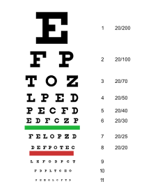
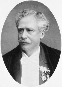
The first person to understand the need for a standardized way of measuring vision was a German ophthalmologist named Heinrich Keuchler.  Around 1842, Keuchler created the very first visual acuity chart.  Actually, he created 3 different ones to prevent his patients from memorizing the letters… you all know who you are.

A few years later in 1862 the vision chart was refined even further by Herman Snellen (pictured right) who created what he called optotypes.  Optotypes are simply specifically sized characters printed in a grid form.  This ingenious decision to start using standard font sizes of the time made these charts easily reproducible and they quickly became the global standard.

So popular was this invention, that the vision chart you read at the doctor’s office today still bears his name and the Snellen Chart (above) has sold more copies since it’s invention than any other poster in history!

Another fun fact about the Snellen and other vision charts is that the letters G, I, J, M, Q, W, X, and Y never appear.  See, reading the chart isn’t that hard, I just made it even easier!


What do the numbers in 20/20 stand for?


When Snellen first designed his reading chart, his optotype characters were meant to be read from 20 feet and if you could, you were considered 20/20.  Therefore, the first 20 in 20/20 simply stands for 20 feet.  This distance is important because past 20 feet the focusing of our eyes is the same as it is for any distance further out.  This distance is called “optical infinity” and is the distance that vision is measured.  In the past, it wasn’t uncommon to be examined in 20 foot long exam lanes.  These days however, it is far more common to use electronic charts or to incorporate mirrors to make the Snellen chart seems further away.

The second 20 in 20/20 is a little trickier to understand, but it basically compares how well you can see the chart compared to a normal 20/20 person.

For example, what if your vision is not the perfect 20/20 but only 20/40?

Well, this means that the smallest line of letters you can read at 20 feet is the same as for someone with normal vision who is 40 feet away.  And, if you could only see the 20/100 line of letters at the standard 20 feet, a normal person could see those same letters from 100 feet away.  This system is the same for every other measurement in between like 20/30, 20/80, 20/200, etc.

It also works the other way.  If you are blessed enough to have better than normal vision at 20/15 or even 20/10 vision, then you can see at 20 feet what a normal person can only see at 15 or 10 feet.


Best Corrected Vision


The last thing that it is important to discuss when it comes to visual acuity is the concept of Best Corrected Vision.  Best corrected vision is the very best that you can see when you have the best glasses or contact lens prescription possible.  This is typically the way that visual ability is described.

Therefore, the question of, “What is my vision without glasses” really isn’t important.  If you take your glasses off and can barely see the “Big E” on the Snellen Chart (which is 20/200) but with glasses you easily read the 20/20 line – you are not legally blind, your vision is considered the normal 20/20.


I hope this has cleared up some confusion around visual acuity, but if you have any additional questions or topics for future discussion, please contact me at [email protected].

We love to educate!

Thanks again for stopping by and take care of those eyeballs!!

  • Nick Wolf, OD

What does it mean to be Color Blind?

Color Blind is a term that is often thrown around and usually with negative connotations.  But for someone who is color blind, what does it actually mean and how did it happen?  What does it mean if your child is diagnosed with color deficiency?  What does the world look like to someone who is color blind?

All great questions we will answer today on Eye to the Future.

What is Color Blind?

eye dilationBefore we get into what it means to be color blind or color deficient, let’s first look at how we actually see color.  As discussed in a past blog post on how animals see in the dark, there are two types of light sensing cells in the retina: Rods and Cones.  Rod cells in the retina are good at sensing movement and work well under dim illumination.  Cones cells, by contrast, need lots of light to work properly, but are capable of discerning incredible detail as well as sensing color.

What was not discussed before, is that there are actually three different types of cones cells in the human eye that each sense different wavelengths of light: one type that senses the color Green, one for Red, and one for Blue.  When light enters the eye, these three cells are activated to varying degrees based on the color or wavelength of the light.

Astonishingly, with just these three different color sensing cones types, the human eye is able to distinguish over 7 MILLION different colors!!  Now that’s pretty cool.

But what happens if one, two, or even all three of these color cells are either not working properly or completely absent?  The result is color deficiency or color blindness.

Types of Color Blindness

There are two forms of color deficiency: acquired and hereditary (i.e. genetic).  Acquired color blindness occurs from some uncommon eye diseases or as a rare side effect of some medications, however, today we will discuss the far more common hereditary form.

Hereditary color deficiency is very common and has a strong male tendency, afflicting just over 1 in 12 males but only 1 in 200 females.  Females are much more likely to “carry” the gene for color deficiency and pass it to their sons, whereas males are more likely to “express” the condition and actually be color blind themselves (more detailed explanation).

Many people believe that color blindness means a total lack of color vision.   True color blindness can occur when just one of the three color sensing cells develops: this condition is called monochromacy.  In actuality though, this condition is exceedingly rare (about 1 in 1 million new births).

Ironically enough, the vast majority of “color blind” individuals actually do see color.  However, they see color differently because they only have two of the three cone types (suffix –anopia) or they have all three cones cell types but one of them is not functioning properly (suffix –anomaly).  These conditions are named for what color cell type is defective and statistically it is most often the Green color cone cell type that is affected.  As you can see below, if any cone cell type is missing or ineffective it has a dramatic effect on the way color is perceived.

Normal Vision

normal color

Protanopia or Protanomaly – Red Cone Cells absent or defective

protanopia

Deuteranopia or Deuteranomaly – Green Cone Cells absent or defective

deuteranopia

Tritanopia or Tritanomaly – Blue Cone Cells absent or defective

tritanopia

Diagnosis, Treatment, and Impact of Color Deficiency

Color Blind
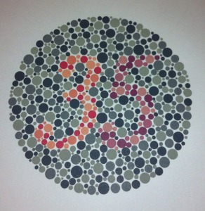
Diagnosis of color deficiency is very straightforward during a normal eye exam.  This is done by using colored plates with dots in various patterns to spell out letters, numbers, lines, or shapes.  These color plates (like the Ishihara plates at right) use specific colors chosen to be difficult for color deficient individuals to tell the difference.  As a result, these individuals have trouble identifying the number or numbers on the plate, but for a color normal individual this will be very easy.

Considering that color vision defects are mostly a generic/inherited condition, there is really no effective treatment to regrow or fix cone cells that are either absent or not working properly from birth.  The good news is that most of the time being color blind or color deficient has very little impact on an individual’s day to day lives.  There are some professions that simply require normal color vision like an artist or painter, airplane pilot, policeman or fireman, as well as licensing for some commercial vehicles.  However, for the most part, being color blind or color deficient, is quite common and does not prevent normal eye development, academic achievement, or professional success.

Aside from some occasional gentle ribbing by a spouse or coworker about a questionable attire selection, these individuals live normal and colorful lives.

As always, thank you for stopping by Eye to the Future and keep those questions coming!

Take care of those eyeballs!

– Dr. Wolf

For Loads more great information on Color Deficiency as well as some shocking examples of vision with color deficiency please visit the Colour Blind Awareness website.

What is eye dilation and why do I need it?

As I’m sure you can imagine, as eye care professionals we get asked about eye dilation A LOT!  Although patients are frequently aware that eye dilation is part of a comprehensive eye exam, they often use the side effects of dilation as a reason for not doing it – we’re all friends here, you can admit to this tactic.

This article will try to convince you of the importance of eye dilation by exploring what eye dilation actually is as well as how the benefits of this simple test can save eye sight.  We will also dispel some eye dilation myths and misconceptions.

The Anatomy of Eye Dilation

The reason that eye dilation is even necessary is because of the anatomy of the human eye.  Assessing the health of the front of the eye is a fairly easy task using our light microscopes; however, viewing the inside of the eye is a challenge.  When light enters the eye through the pupil, there is a pretty nifty little neurological loop from your eyes to something called the Edinger-Westfall nucleus in your brain.  This part of the brain links with the muscles of your iris to constantly control the size of the pupil and thus the amount of light alloweye dilation
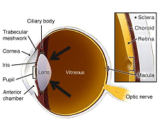
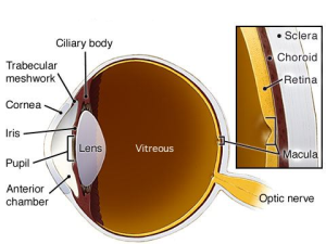
ed into the eye.  Being in dimmer environments makes the pupil open wider to let light in to improve vision, while being in brighter environments causes the pupil to constrict to decrease light sensitivity and glare.  This is a great system for humans but a terrible system for eye doctors because the very bright lights from our microscopes makes the pupil so small we can’t see through it!  It’s like trying to look through a keyhole into a room and reduces our view of the inside of the eye to only about 10-15%!!  Going to an eye doctor and NOT getting your eyes dilated it like getting a physical but only letting your primary care doctor look at one leg.

Eye Dilation for Complete Eye and Systemic Health

In truth, eye dilation on adults is done to make sure ALL parts of your eyes are healthy and at Precision Family Eye Care, we would argue that eye dilation is the MOST important part of a comprehensive exam.  By dilating the eyes we are able to see 100% of the interior of the eye and rule out all eye diseases: from Glaucoma and Macular Degeneration, to Cataracts and even eye tumors.

BRAOThe benefits of eye dilation don’t stop with the eye.  The interior of the eye is unique in that is it the only place in the body where nerve tissue and blood vessels can be viewed without any advanced procedures or expensive testing.  This allows your eye doctor a perfect opportunity to use the information gleaned from your dilated exam to identify problems and conditions elsewhere in the body.  There are quite literally hundreds of systemic conditions (the short list) that can result in changes to the interior of the eye but some of the more common conditions are Diabetes and cardiovascular disease.  The photo at right is a great example.  This patient had no complaints, but a dilated exam found a cholesterol plaque in a retinal artery.  This patient needs emergent care and testing due to a high stroke risk.  At Precision Family Eye Care, we work closely with doctors from all specialties on cases like this to communicate eye health findings and improve care.

Side Effects and Myths about Eye Dilation

First of all, it is important to note that there is NO substitute for dilation, although some offices may try to sell you alternatives.  The newer digital systems these companies offer can take relatively good pictures of the retina.  The problem is that these photos are only able to image about 65-70% of the retina and the 2-dimensional picture with poor resolution is not adequate to diagnose most eye diseases.  Simply put, this technology does not reach the standard of care needed for a Comprehensive eye exam AND this testing is an additional fee whereas eye dilation is an included service with any visit.

With eye dilation, there are some temporary side effects.  The most obvious side effect is light sensitivity because the larger pupils allow in more light.  This is easily solved with sunglasses and temporary shades can be supplied by your eye doctor.  Secondly, if you are under 50 years of age, the dilation drops also affect your ability to focus on up-close objects.  These two side effects are temporary and only last 2-4 hours.  Dilation will not affect your distance vision to the point where you will have trouble driving a car; a dedicated driver is an option but is certainly not a necessity.

How often should eyes be dilated?

Eye dilation is necessary at every visit for patients with certain systemic diseases like diabetes or in patients with confirmed eye disease.  The vast majority of early staged eye diseases do not cause obvious symptoms (like blurry vision).  Therefore it is necessary for patients to received routine dilation to rule out disease every couple years up to the age of 50 after which yearly dilated exams are strongly recommended.

Eye dilation definitely falls under the category of “necessary evil” but we hope that this discussion has convinced you of the importance of this procedure.

If it has been a few years since you have had your eyes dilated, we strongly recommend that you schedule a visit with an eye care professional.

See you next time on Eye to the Future and remember to take care of those eyeballs!

– Dr. Wolf

October: Eye Injury Prevention Month

rust ring
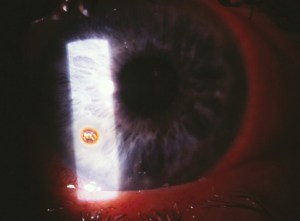
A friendly gentlemen came in to see me today with a very angry eye and reported that he had been working on his son’s car earlier in the week and his eye has been red and scratchy every since.  As I was preparing to drill out the large hunk of rusted metal (left), it dawned on me, October is Eye Injury Prevention Month!

Statistically speaking, over 2.5 million Americans will experience an eye injury this year.  Some injuries are simply unavoidable or arise from falls or motor vehicle accidents, most injuries occur due to negligence and a lack of proper protective eye wear.

It is a common misperception that all eye injuries occur “on the job.”  In reality, about 45% of eye injuries actually occur at home and (I’m sorry guys) men have much higher risk for eye injuries than women.

Common places/tasks for Eye Injury

  • Yardwork

    Mowing the lawn or being outside while others are mowing.  This is doubly true for power edging or using gas or electric powered trimmers.  Rocks, organic debris, and dirt can be picked up and propelled with dangerous speed by all of these machines and tools.

  • corneal abrasionWorkshop

    As a carpenter I can assure you, the force with which a power tool expels projectiles can be simply staggering (picture right is a deep corneal laceration from  metal lathe work).  Think about the work that needs to be accomplished and which tools you will be utilizing and plan accordingly.  Also consider what chemicals/stains/sprays/fumes your eyes may come in contact with.

  • Working on the car

    There’s more at risk here than just a bruised knuckle.  Working on and especially under a car is a major source of metallic foreign bodies.  Additionally, arcs, sparks, and battery acid can be blinding.  Keep protective goggles in your garage and vehicle and wear them for any and all repairs.

  • Housework

    If you actually read the ingredients of most household cleaners, you may be surprised.  Make sure these dangerous chemicals, cleaners, and disinfectants are used in a well-ventilated area and with proper eye protection.  Chemical splashes from some of these hazardous cleaners can cause permanent burns and vision loss.

In order to combat these potentially blinding situations, we strongly recommend that all households have at least one pair of ANSI-approved safety glasses to use for these tasks to reduce the chance of eye injury.  In order for glasses to be marketed for safety purposes the lens and/or frame must bear the required marking of ANSI Z87 or preferably Z87+ (the ‘+’ denotes high impact resistance).  Protective eyewear of this kind is readily available at all hardware stores or purchased from your local eye doctor.  It is important that safety glasses fit close to the face with minimal gaps for optimum protection against chemical splashes.  Click here for additional information on safety eye wear.

If a chemical splash does occur, rinse the eye with copious amounts of luke-warm tap water for 15 minutes.  That may seem like a long time but this is imperative.  The sooner this gets done the better, as this will dilute and neutralize the caustic compounds.  Then seek treatment from your local eye care provider.

The cornea is extremely sensitive and has more nerve endings than any other tissue in the body.  Anyone who’s had an eye injury can tell you; they hurt… A LOT!

Accidents happen, but Ben Franklin said it best, “an ounce of prevention is worth a pound of cure.”

We hope you have enjoyed this quick talk on eye injury prevention.  If you have any additional questions or think of a topic for future discussion; drop me a line at [email protected].

Wear your safety glasses and take care of those eyeballs!

STUDY: Contact Lenses in Pediatrics (CLIP) Study

Contact Lens Fitting

Contact Lenses in Pediatrics

As eye care professionals, it is difficult to count the number of times this scenario has played out in our exam rooms.

A child presents for their back to school eye exam with a parent in tow.  The exam is uneventful, the prescription is checked, the eye health is thoroughly evaluated, and after all testing is completed and the findings are conveyed to the parent or caregiver, the real crux of the visit finally reveals itself… someone brings up contact lenses.

Suddenly, what was a cordial visit gets a bit more heated.

Parent:  “You are WAY too young! I didn’t get them until I was in highschool!”

Child: “But my friend Billy got them two years ago!”

Parent:  “Maybe if you could keep your room clean, your mother and I would consider it!”

Child: “But I don’t like wearing glasses, especially under my baseball helmet!”

Parent:  “It isn’t safe at your age, we’ll talk about it next year when you’re 13.”

This usually continues rapid fire for 15 to 20 seconds until, exasperated after having made their cases, both parent and child turn in anticipation toward me to cast the deciding vote…

“In reality, you are both right,” I will undoubtedly say.

Let’s look at this rationale in two ways, first by looking at an actual study on the matter and secondly by looking at contact lenses in pediatrics from a real world view point.

The Contact Lenses in Pediatrics or CLIP Study

This study sought to answer the age old question of when is it appropriate (i.e. safe) for children to begin wearing contact lenses?  This large study enrolled 84 children from the ages of 8 to 12 along with 85 teens from the ages of 13 to 17 and all participants were brand new to contact lens wear.  The demographics of the participants in this study were reflective of the general US population in terms of gender and race.  All participants where properly trained, fit with the same brand of contacts for continuity, and then followed at one week, one month, and 3 month intervals, when problems from new contact lens wear are the highest.  The follow up visits looked for problems like reduced vision, trouble with putting in and taking out contacts, proper fitting of the contact, and checking the health of the ocular surface with a microscope for signs of inflammation or infection.

When all the data was tabulated, there was no statistical difference in the safety or ability of contact lens wearers in either of the two groups.  To put it another way, if proper contact lens training and fitting was performed by the doctors and staff, then 8 year old children wore contact lenses as safely and successfully as 17 year old teens!

Not only that, another arm of the CLIP study looked at the child’s perceived “quality of life.”  This survey showed a remarkable 24% improvement in children’s perception of contacts compared to glasses.  The largest areas of contact lens preference came in the categories of overall appearance, improved vision, more comfortable activities (sports), and reported self esteem.  In other words, all the things that adults love about wearing contacts… children and teens did as well!!

In Reality

Although the studies clearly show that children and young adults of a wide age range can wear contact lenses safely and enjoyed doing so, it certainly doesn’t mean that all children and young adults should wear contacts.  The Fairness to Contact Lens Consumer Act (FCLCA) was signed into law in 2003 (way back when congress actually passed legislation), and declared contact lenses as “medical devices,” and for good reason!!  Any eye care professional has a slew of horror stories and will willingly tell you that rarely does a day go by without a contact lens wearer presenting to the office for some problem directly related to contact lens over-wear and abuse.

Not surprisingly, severe complications from contact lens wear are a significant contributor to avoidable and preventable blindness in the US.  Therefore, far more than any age requirement for contact lenses, it is imperative that there be a responsibility and maturity requirement and especially when you are dealing with the eyes of a child.

When the parents feel that a child has demonstrated the responsibility level required do all the things necessary for proper and safe contact lens wear; no overnight wear, proper care and cleaning, on time replacement, etc. The studies clearly show that with proper training and doctor skill, contact lens wear in children and young adults is a safe, appropriate, and patient appreciated alternative to spectacles.

Thank you as always for stopping by Eye to the Future; we hope you have enjoyed this discussion.  Please feel free to email any questions or topics for future discussion to [email protected].

Take care of those eyeballs and we’ll see you next time!

CLIP Study

Back to School: Don’t forget the Eye Check!

PediatricEyeCareWith school back in full swing, Precision Family Eye Care and the Eye to the Future blog want to take a moment this week to impress upon all parents the importance of getting routine vision exams for all school age children.

School and Pediatrician screenings are simply not adequate, and unlike adults, children can’t articulate their vision problems because they don’t understand that the way they see the world isn’t “normal.”  Uncorrected prescription (like nearsighted, farsighted, and astigmatism) as well as eye alignment and focusing problems, and even ocular diseases can all inhibit the ability of your child to use their eyes properly.  This leads to disrupted vision and potentially vision threatening conditions being simply overlooked or even causing learning problems and leading to a diagnosis of LD (Learning Disorders), ADHD or even dyslexia.

The learning environment today is flush with visual tasks: smart boards, iPads, digital learning, computer work, homework, test taking, etc.  It shouldn’t surprise us then, that learning experts estimate that roughly 80% of all learning is dependent upon the sense of sight and unfortunately, at least 25% of school aged children have a visual impairment that may prevent a child from reaching their full academic potential.

Please make sure that your child has the proper tools for academic success this year by scheduling a comprehensive exam with your eye doctor today!

Patient Question: “How do animals see in the dark?”

As is often the case, most of the ideas for this blog come from actual questions asked by patients.  On Eye to the Future today we will be answering one of these questions I received just last week, “How do animals see in the dark?”

GREAT QUESTION!

How exactly does my dog and cat run scot-free through a dark room but I stub my toes and step painfully on toys?

The answer: it’s all in the design of the eye.

Thanks for stopping by, next week on Eye to the Future we will be discussing… kidding of course.

To answer this question fully, there are actually three major anatomical differences between the human eye and animal eyes capable of great night vision.

Rods versus Cones

CataractsDiagramAs most are aware, the retina is the covering of the inside of the eyeball.  Although this thin tissue is only about 3 human hairs thick, it is essentially the ‘film in the camera’ and is responsible for sensing the light that enters the eye and transmits that signal back to the brain where it is processed.  The actual cells that sense the light are called photoreceptors and there are two main types: rods and cones.  Although they both are important in seeing, they do so very, very differently.

Cones are responsible for color vision and high acuity vision however these cells only function in bright light.  Rod cells, by contrast, are much less sensitive to colors and fine details but are able to detect light and function in much lower light situations.

In humans, 90% of our cones cells are located in the central part of our vision which allows us vivid, clear, and colorful central vision, but general poor vision under scotopic (dark) conditions because of a lack of rods in our central vision (graph at right).  Rod and Cone DistributionBy contrast, nocturnal animals have retinas composed mostly or even entirely of rods cells, especially in their central vision.  This affords nocturnal animals great night vision; however, their lack of cone photoreceptors leaves these animals with little to no color vision and an overall decrease in clarity compared to humans.

Bigger is Better

Since nocturnal animals cannot benefit from the crisp vision that cone cells allow, they require a much larger image to focus on their retinas.  The easiest way to accomplish this is with a larger eye and larger eye structures (like a stronger focusing cornea and lens as well as a larger pupil to let in more light).  The human eye is about 24 mm (about 1 inch) in diameter.  The eyes of nocturnal animals are substantially larger even through the animal itself can be much, much smaller.  The owl is a perfect example of this since it has an eye that is about the size of a human eye but gives enough visual information to allow the owl to be a successful night bird of prey.  However if we are talking size, the most dramatic example and clear winner of the “Biggest Eye on the Planet” award goes to the giant squid which is able to visualize prey in the pitch black of the ocean’s depths with an eye that’s the size of a dinner plate!!

Tapedum Lucidum

Although larger eyes and specialized retinal cells are certainly useful, nocturnal animals have one final trick up their sleeves to help with night vision.  The retina in all animals is a clear, see through tissue.  The reason for this is because the rod and cone cells that actually sense the light are at the back of the retina, so the layers above need to be clear to let light get through.  Now in a human eye, once the light goes through the retina it hits the retinal pigmented epithelium or RPE.  This thin layer of cells is highly pigmented and absorbs excess light that gets through the retina to reduce glare/scattered light and improve overall clarity of vision.

However, many nocturnal animals like deer, raccoons, cows, horses, and even dogs and cats, have developed a specialized RPE behind the retina called the Tapedum Lucidum that does the exact opposite.  Instead of being highly pigmented to absorb the light that the retina does not collect, the Tapedum Lucidum is a highly reflective tissue, like a mirror, that actually bounces the light back through the retina for a second chance to be detected.  This adaptation effectively doubles the chance that even very low amounts of light will be detected by the retina.  Reflections off of this tin foil like Tapedum Lucidum is also what causes the brilliant white reflections from the eyes of nocturnal creatures, like a deer in headlights or even a photo of the family pet.

Tapedum LucidumWe hope you have enjoyed this long answer to a short question.  Next time on Eye to the Future, we will be discussing polarized lens.  How they work, how they’re made, and why they are so, so useful for clarity and vision.

I’ll leave you with a picture of our sweet boy, Remy, taking a rest from reeking havoc and showing off his Tapedum Lucidums.

See you next time on Eye to the Future.

STUDY: Age Related Eye Disease Study (AREDS 1 & 2)

On occasion, there are landmark studies regarding the eyes that everyone should know about.  Today on Eye to the Future we will be discussing two such studies, by far the largest and most important studies on preventing the progression of the aging condition Macular Degeneration.  (For those not interested in the science, the results and take home points are summarized at the bottom of this post.)

Dry Macular DegenerationAge Related Macular Degeneration (ARMD or simply AMD) is the leading cause of new blindness in the United States for individuals over 65 years old.  This condition is not a new problem, however, for many decades the mechanism for why this damage occurred was unknown and treatment options for this condition were woefully limited and relatively ineffective.  Thankfully, in the last decade and a half, some genius innovations in treatment, as well as some landmark studies on prevention, have given individuals afflicted with this blinding disease a fighting chance at retaining vision throughout their lives.

Wet Macular DegenerationMacular Degeneration can be classified into two categories Dry and Wet (for an in-depth discussion on the difference between the two, please view our website for a thorough discussion).  Dry Macular Degeneration (above) always comes first and by itself can cause vision loss, however, more often it is the 10-15% of patients that convert to the Wet form (with its associated bleeding and scarring) that suffer severe permanent damage.  Therefore, researchers set out to look for ways at preventing or at least slowing down the conversion to this more aggressively blinding Wet form (right).  The research culminated in the Age Related Eye Disease Study (AREDS) that was first published in October of 2001, and overnight, changed the way eye care practitioners approached macular degeneration.

AREDS 

It had been hinted at in smaller previous studies that antioxidants as well as the trace element Zinc appeared to be helpful for macular degeneration patients.  As a direct result, many patients starting taking high doses of these vitamins and minerals without any set dosing amounts, schedules, or any singular study that ensured this practice was even effective… or safe for that matter!!  It became clear that a definitive study was needed and the National Eye Institute, one of the government’s Institutes of Health, brought onboard a decorated panel of eye doctors, biochemists, and nutritionists who came together and concocted the follow mixture of supplements.

Vitamins

o   Beta-Carotene (15mg) which is converted in the body to Vitamin A

o   Vitamin C (500 mg)

o   Vitamin E (400 IU)

Trace Minerals

o   Zinc (80 mg), which was shown to be effective in earlier studies

o   Copper (2 mg) to prevent anemia while taking high doses of Zinc.

Over 3500 patients with various amounts of Macular Degeneration took this supplement for a minimum of 5 years.  Once all the data was tabulated, the most encouraging results were for the patients who needed it most with intermediate to advanced Macular Degeneration.  The results for patients in this category are outlined below.

Vitamins Alone

  • Reduced the risk of advanced ARMD by 17%
  • Reduced vision loss by 10%
Trace Minerals Alone

  • Reduced the risk of advanced ARMD by 21%
  • Reduced vision loss by 11%
Vitamins AND Trace Minerals

  • Reduced the risk of advanced ARMD by 25%
  • Reduced vision loss by 19%

Once it was shown that this nutritional supplement formula of both vitamins and trace minerals decreased advanced Macular Degeneration by 25% and vision loss by nearly 20%, many different nutraceutical companies began reproducing the AREDS Formula with brand names that you have likely heard of or have seen in stores like I-Caps™, PreserVision™, Ocuvite™, et al.

AREDS 2

Not long after the AREDS was published, the researchers behind the AREDS study went back to work on an even larger more detailed study using the information gleaned from the first study.  The results of the AREDS 2 were just released in 2013.  This improved study adjusted two of the past components of the AREDS formula and also added a third.

  1. Beta-Carotene:  Very high doses of Beta-Carotene/Vitamin A supplements have been linked to an increased risk of Lung Cancer in current or former smokers.  Since smoking in-and-of-itself is already a significant risk factor for developing Macular Degeneration, beta-carotene was removed completely for safety reasons and replaced with two other carotenoids: Lutein (10 mg) and Zeaxanthin (2 mg) which were deemed a safer and equally effective substitute for Vitamin A.
  2. Zinc:  It was suspected that high dosages of Zinc are linked to digestive problems.  Reducing Zinc (to 25mg)  showed the same benefit as the AREDS formula, but removing Zinc from the formula completely was detrimental.
  3. Omega 3 fatty acids (fish oils):  These compounds are a known anti-inflammatory and antioxidant.  Researchers added this compound to the AREDS formula in hopes of additional reduction in risk, however, Omega 3’s were shown to have no effect on Macular Degeneration progression.

AREDS 1 and AREDS 2: Take Home Points

  • Any patient with confirmed macular degeneration will benefit from eye specific supplements, but these supplements are most helpful for patients with moderate to advanced Macular Degeneration.
  • These supplements should be taken IN ADDITION to any multivitamins, not as a substitute (9 out 10 patients in the AREDS 2 study were also taking a concurrent multivitamin).  If only one supplement is desired, there are formulations of these supplements that combine the AREDS formula with a multivitamin for simplification.
  • The original AREDS formula is fine for many, if not most patients.
  • Any smoker or former smoker should avoid beta-carotene supplements completely and use only the AREDS 2 formula with Lutein and Zeaxanthin instead.
  • Omega 3 fatty acids (fish oils) although wonderful for the body and the eyes, did not have a meaningful impact on Macular Degeneration.
  • These studies ONLY found benefit for patients with diagnosed Macular Degeneration.  These supplements are not designed, and should not be taken for “routine” eye health or even for patients with a family history of Macular Degeneration.

We hope you found this discussion interesting.  As always, if you have any questions or have ideas for future topics feel free to contact us and ask.  We love questions!

Eye to the Future next week will be a fun one… but I’ll let it be a surprise.


“Pink Eye” Myths: What it is and What it’s not.

Well, it’s back to school time and that means it’s also time for some germs to be spread and flus to be caught.  This time on Eye to the Future we will be talking about the eye’s contribution to the germ problem with Pink Eye.  The term Pink Eye gets batted around quite a bit in most people’s lexicon.  The problem with this term is that is really doesn’t describe what the problem really is or how a patient’s irritated eye should be monitor/treated or if that patient has the possibility of spreading their eye condition to others.  Therefore, let’s dispel some myths and define some terms.

Conjunctivitis, commonly referred to as Pink Eye, is the technical (but nonspecific term) for any inflammation or infection of the conjunctiva.  The conjunctiva is a thin, clear, slippery tissue that covers the white of the eye and inside of the eyelids.  This tissue is the first line of defense and acts as an overseer against any viruses, microbes, or irritants that get into the eye.  When the conjunctiva senses that the environment on the front of the eye is not healthy or a potential for infection exists, it calls in reinforcements from the rest of the immune system for help and makes the eye look red or pink.  It is important to note that this reaction happens not only against infections, but in ALL instances of eye irritation.

Therefore, a red or pink eye does not mean that you have Pink Eye, or even a conjunctivitis for that matter, it just means that the eye is unhappy for some reason.

If the diagnosis is in fact conjunctivitis, there are three different types of conjunctivitis: allergic, bacterial, or viral conjunctivitis.  Pink Eye is widely considered to be ONLY the viral type.

–        Allergic conjunctivitis is an extremely common occurrence especially around here in the Ohio River Valley.  As the name implies, this is an allergic reaction that happens due to pollen, dust, dander, contact lenses or chemicals (like soaps or makeup).  Commonly this condition presents in both eyes with watering, itching, and a “soggy” eye appearance.  Treatment here is to quell the inflammation and this is accomplished best with prescription eye drops.  Since there is an environmental cause, this conjunctivitis is not contagious.

–        Bacterial conjunctivitis is actually rather uncommon.  True bacterial infections of the conjunctiva present with a sudden swollen, very red eye along with a heavy mucus discharge.  Antibiotics are often given as treatment for conjunctivitis even though a bacterial infection is rarely the cause.  This form can be contagious when a mucous discharge is still present.

–        Viral Conjunctivitis.  Now, here is the true Pink Eye.  Most conjunctivitis is viral in nature and these infections are spread through direct contact.  The worst of these is the adenovirus which quickly spreads through workplaces, households, and schools.  The adenovirus has a latent period of 7-14 days from initial contact with the virus to the start of eye symptoms developing.  Symptoms include redness, watering, itching, and tenderness of the preauricular lymph nodes (the ones just in front of your ear).  Symptoms start in one eye and often spread to the other eye in 3-5 days and failure to seek treatment could result in scarring of the cornea and the possibility of permanently decreased vision.  Treatment for most viral conjunctivitis involve prescription antiviral eye drops and/or in office treatment.  It is EXTREMELY important with ALL viral conjunctivitis to wash your hands frequently and to stay away from work or school until symptoms improve.

We hope this has shed some light on a common but commonly confusing condition.  It is important to reiterate, that a red eye does not necessary mean Pink Eye or even conjunctivitis.  For any red eye it is important to seek treatment from your eye doctor to get a proper diagnosis and treatment regimen to keep your fellow students, coworkers, and family members safe if the condition is contagious.

Finally… if we are discussing Pink Eye and Myths we should tactfully address one final item.  For the record, performing a certain bodily function on a certain bedding item, while childishly humorous, most certainly DOES NOT spread Pink Eye.

Stay tuned for the next installment of Eye to the Future where we delve into a landmark study on Macular Degeneration that everyone should be aware of: the AREDS (Age Related Eye Disease Study) and how it can prevent blindness.