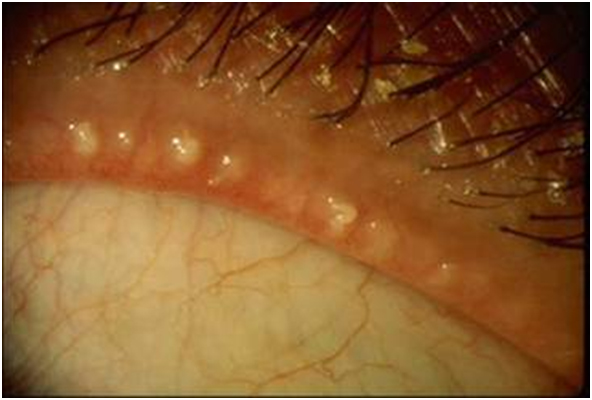
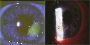
Anterior Uveitis


AnteriorUveitis
Statistics and Associations

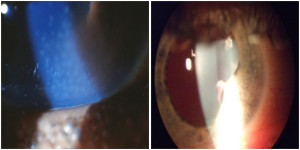
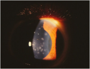
  • Characterized by inflammatory cells and proteins in the anterior chamber of the eye
  • Usually idiopathic
  • Recurrent cases should be evaluated with lab tests for autoimmune diseases

Management

  • Steroid regimen (dosing is severity dependent) with gradual taper to avoid rebound inflammation
  • Cycloplegic agent to decrease inflammation, improve comfort, and prevent synechia of the iris

Fuchs’ Endothelial Dystrophy


Fuchs-Endothelial-Dystrophy
Statistics and Associations

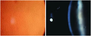
  • Most common hereditary endothelial (deep) corneal dystrophy
  • Strong female predilection
  • Typically develops in fourth to fifth decade of life with gradual progression
  • Leads to decreased, fluctuating vision usually worse in the morning
  • Vision decrease secondary to corneal swelling

Management

  • Generally no treatment until substantial vision decrease
  • Endothelial keratoplasty is the main surgical intervention

Epithelial Basement Membrane Dystrophy (EBMD)


Epithelial-Basement-Membrane-Dystrophy-EBMD
Statistics and Associations

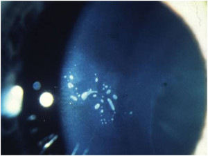
  • Most common hereditary corneal dystrophy
  • Bilateral with early onset (age 30); slight female predilection
  • May lead to scarring and decreased vision in severe cases
  • 10% of patients with EBMD have corneal erosions and 50% of recurrent corneal erosion patients have EBMD

Management

  • Generally no treatment, unless recurrent erosion develops
  • May consider artificial tears and/or hypertonic ointment at night to decrease risk of erosions

Band Keratopathy


Band-Keratopathy
Statistics and Associations

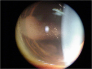
  • Superficial calcification of cornea
  • Associated with chronic corneal irritation, gout, and hypercalcemia

Management

  • No treatment recommended until visually significant
  • EDTA chelation, superficial keratectomy, and penetrating keratoplasty all common surgical interventions

Herpes Simplex Keratitis


Herpes-Simplex-Keratitis
Statistics and Associations

  • Most common cause of central infectious keratitis
  • Occurs secondary to reactivation of HSV type I through nervous system
  • Following one HSK infection, 25% chance of recurrence in one year; 50% chance of recurrence in two years
  • Suppressive therapy reduces risk of recurrent keratitis by almost 50%

Management

  • Topical antiviral (Viroptic™ q2h or Zirgan™ 5xday) for two weeks, oral antiviral (valacyclovir 500 mg PO TID) for one week.
  • Consider suppressive therapy (valacyclovir 500 mg QD) for recurrent cases

Corneal Ulcer


Corneal-Ulcer
Statistics and Associations

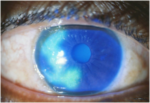
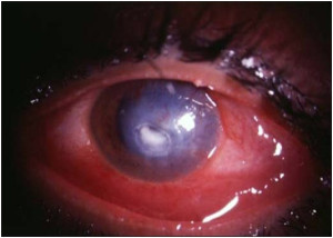
  • Destruction of corneal tissue with strong association to contact lens overwear as well as dry eye and mechanical trauma
  • If central, strong probability of decreased vision post treatment secondary to scarring

Management

  • Discontinue contact lenses
  • Determine etiology and treat aggressively with strong antibiotics with steroids later to decrease scarring, earlier with central ulcer to increase final visual outcome

Subepithelial Infiltrates


Subepithelial-Infiltrates
Statistics and Associations

  • Strong association with contact lens overwear and overnight wear
  • Usually peripheral with increased risk of overlying ulcer formation
  • Can be sterile or infectious

Management

  • Discontinue contact lens until resolution
  • Treat as infectious with broad spectrum antibiotic every hour then taper, steroid effective in resolution and preventing scarring

Neovascularization/Pannus


Neovascularization-Pannus
Statistics and Associations

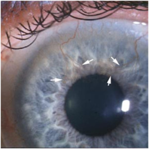
  • Micropannus (< 2mm) common with contact lens wear/overwear secondary to corneal hypoxia
  • Pannus (> 2mm) can decrease vision secondary to scarring and stromal hemorrhage
  • Also common with dry eye, ocular rosacea, and chemical burn

Management

  • Discontinue contact lens wear and fit with higher oxygen lens
  • Treat any underlying source of ocular inflammation

Recurrent Epithelial Erosions (REE)


Blepharitis Meibomitis
Statistics and Associations

  • 50% association with anterior basement membrane dystrophies
  • Also common following severe corneal abrasions; especially from sharp objects such as a fingernail or paper

Management

  • Same as corneal abrasion until epithelium is healed, then add hypertonic ointment
  • Doxycycline oral medication for persistent cases
  • Consider debridement in cases of multiple reoccurrences

Corneal Abrasion/Foreign Body


CornealAbrasionForeignBody
Statistics and Associations

  • Abrasions usually trauma related
  • Foreign body usually metal, glass, or organic material

Management

  • Removal of foreign body, antibiotic coverage, cycloplegic if needed, and bandage contact lens for comfort
  • Follow daily until wound heals
  • Consider hypertonic (salt) solutions or ointment to decrease risk of recurrent epithelial erosions